Conditions · Surgery and trauma
Midface fractures (Le Fort, zygomatic, nasal) !
Midface fractures break the bones between the eyes and the upper teeth. They can block the airway, bleed heavily, and affect vision and the bite.
Updated 2026-11-01
Structures involved
Tap one to see what it is and open it in 3D.
- Maxillae
- Zygomatic bones
- Nasal bones
- Maxillary sinus
- Sphenoid bone
- Ethmoid bone
- Nasal septum
- Nasal cartilages
- V2 maxillary
- Palatine bones
- Frontal bone
- Tear drainage
- Mandible
What it is
Fractures of the middle third of the facial skeleton: the maxillae, zygomatic bones, nasal bones, the orbits and the naso-orbito-ethmoid area. Key patterns are nasal fractures (the most common facial fracture), zygomaticomaxillary complex (ZMC, 'tripod') fractures, and the Le Fort I, II and III fractures, which separate part or all of the midface from the skull base. All Le Fort fractures involve the pterygoid plates of the sphenoid bone.
Who gets it
Facial fractures mainly affect young adult men. In Colombia and Latin America the leading causes are interpersonal violence, motorcycle crashes (often without a full-face helmet), falls and sports. Nasal fractures make up roughly half of facial fractures; true pure Le Fort patterns are less common than mixed fractures.
Causes
- Assault and interpersonal violence
- Motorcycle and car crashes
- Falls, including falls under the influence of alcohol
- Sports injuries
- Work-related and gunshot injuries
Risk factors
- Young male sex
- Alcohol and drug use
- Riding a motorcycle without a full-face helmet or not wearing a seatbelt
- Contact sports without facial protection
- Osteoporosis in older adults who fall
Pathophysiology
The midface is built of thin bone reinforced by vertical and horizontal buttresses that absorb forces from chewing. A blow overwhelms these buttresses along predictable weak lines. Le Fort I separates the tooth-bearing maxilla horizontally; Le Fort II forms a pyramid that includes the nasal bones and the orbital rim; Le Fort III separates the whole midface from the cranium. Fractures through the orbit and ethmoid can entrap eye muscles, injure the infraorbital nerve or tear the dura and cause a CSF leak. The face has a rich blood supply, so bleeding into the nose and mouth can threaten the airway.
Classification
- Nasal bone and septal fractures
- Zygomaticomaxillary complex (tripod) fracture: zygomatic arch, frontozygomatic suture, infraorbital rim and orbital floor, and the maxillary buttress
- Isolated zygomatic arch fracture
- Le Fort I (horizontal, 'floating palate')
- Le Fort II (pyramidal, 'floating maxilla')
- Le Fort III (craniofacial disjunction, 'floating face')
- Naso-orbito-ethmoid (NOE) fractures
Symptoms and signs
- Facial swelling, bruising and flattening of the cheek or nose
- Nosebleed and nasal obstruction
- Numbness of the cheek, side of the nose and upper lip (infraorbital nerve)
- Double vision, a sunken eye or bruising around the eyes
- Bite that does not fit (malocclusion) or a mobile upper jaw
- Pain or difficulty opening the mouth (zygomatic arch impinging on the coronoid process)
Diagnosis
- Clinical
- ATLS primary survey first, with cervical spine protection. Then inspect the face for asymmetry, palpate the orbital rims, zygoma and nose for steps, test infraorbital sensation, eye movements, visual acuity and pupils, check occlusion, and grasp the upper incisors and palate to test midface mobility (movement of the palate alone suggests Le Fort I; with the nose, Le Fort II; with the whole face, Le Fort III). Examine the inside of the nose for septal haematoma.
- Tests
- Beta-2 transferrin in nasal fluid if a CSF leak is suspected; blood tests as part of trauma care.
- Imaging
- Non-contrast CT of the face with thin cuts and 3D reconstruction is the standard for all suspected midface fractures except simple nasal fractures, which are usually diagnosed clinically. Include head CT and cervical spine imaging as indicated.
Differential diagnosis
- Soft-tissue facial contusion without fracture
- Mandible fracture
- Isolated orbital floor (blowout) fracture
- Frontal sinus or skull base fracture
- Dental and alveolar fractures
Goal of treatment
Secure the airway and control bleeding, protect vision and the brain, then restore the facial buttresses so that the bite, eye position and facial shape return to normal.
Treatment
- Acute
- Airway first: suction, sitting up if the spine is cleared, and early intubation or a surgical airway when the midface is unstable; nasotracheal tubes and nasogastric tubes are avoided when a skull base fracture is possible. Control nasal bleeding with packing.
- Procedure
- Simple displaced nasal fractures: closed reduction, ideally once swelling has settled within about one to two weeks; immediate drainage of any septal haematoma.
- Surgical
- Displaced ZMC and Le Fort fractures: open reduction and internal fixation with mini-plates along the buttresses, restoring occlusion first; orbital floor reconstruction when needed.
- Medical
- Analgesia, antiemetics, tetanus prophylaxis and antibiotics in selected open or sinus-involving fractures; avoid nose blowing with orbital or sinus fractures.
- Follow-up
- Nursing priorities: airway and breathing, head elevation, repeated visual acuity and pupil checks, watch for clear nasal drainage, neurological observations, mouth care and nutrition with a soft or liquid diet.
Complications
- Airway obstruction and aspiration
- Persistent double vision, enophthalmos or vision loss
- Malocclusion and chewing problems
- CSF leak and meningitis
- Infraorbital numbness
- Facial asymmetry, nasal deformity and obstruction
- Sinusitis and nasolacrimal duct obstruction
Prognosis
Most patients without airway or brain injury do well with timely treatment. Complex Le Fort and NOE fractures may need several operations, and some patients keep numbness, double vision or minor asymmetry.
Prevention
- Full-face helmets for motorcycle riders and seatbelts in cars
- Facial protection in contact sports
- Violence-prevention and alcohol-harm-reduction programmes
Test yourself
Think of the answer, then tap the question to see it.
Which structure is fractured in every Le Fort fracture?
The pterygoid plates of the sphenoid bone.
In a Le Fort III fracture, what moves when the upper incisors are grasped?
The whole midface moves, because it is separated from the cranium (craniofacial disjunction).
Numbness of the cheek and upper lip after a zygomatic fracture suggests injury to which nerve?
The infraorbital nerve, a branch of the maxillary nerve (V2).
Which nasal finding after trauma requires urgent drainage?
A septal haematoma, to prevent cartilage necrosis and saddle nose deformity.
What is the imaging study of choice for suspected Le Fort or zygomatic fractures?
Non-contrast thin-slice CT of the face with 3D reconstruction.
Related conditions
- Nasal fracture and septal haematoma
- Orbital floor (blowout) fracture
- Mandible fracture
- Basilar skull fracture
- Traumatic brain injury
- Epistaxis (nosebleed)
- Ocular trauma: hyphema and open globe injury
- Major trauma (polytrauma): ATLS primary survey
Educational information, not medical advice. For any symptom, see a health professional.
Sources: Mid-facial fractures and their current classification systems: narrative review (Frontiers of Oral and Maxillofacial Medicine, 2025); Facial trauma: MedlinePlus Medical Encyclopedia (MedlinePlus (U.S. National Library of Medicine), 2024).
Spotted a mistake?We are always improving for the community. Tell us what is wrong and our team will review it.Report a mistake
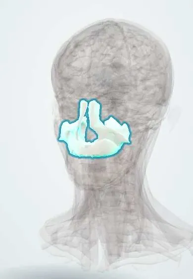
Representative model, for educational purposes only.