Conditions · Infectious and tropical diseases
Actinomycosis !
Actinomycosis is a slow bacterial infection, usually of the jaw, that forms hard lumps and draining tracts and can look like cancer.
Updated 2026-11-01
Structures involved
Tap one to see what it is and open it in 3D.
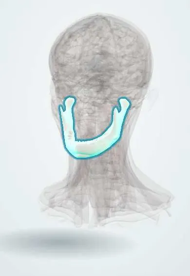
- Mandible
- Masseter
- Submandibular gland
- Gums (gingiva)
- Neck lymph nodes
- Pleura
- Ribs (back part)
- Cecum
- Appendix
- Uterine tubes
- Ovaries
Microorganisms that cause it
Why it happens
- Patterns of acute inflammation: serous, fibrinous, purulent, abscess, ulcer
- Fibrosis in organs: TGF-β and the myofibroblast
What it is
A chronic infection by Actinomyces bacteria that normally live in the mouth, gut and female genital tract. After a break in the mucosa they cause a slowly growing, hard mass that crosses tissue planes and forms abscesses and draining sinus tracts, sometimes releasing yellow "sulphur granules".
Who gets it
It occurs worldwide and is uncommon. Cervicofacial disease is the most frequent form and is linked to poor oral hygiene, dental caries and dental procedures, all common problems in Latin America. Thoracic disease follows aspiration; abdominal disease follows surgery or a perforation; pelvic disease is associated with long-term intrauterine device use.
Causes
- Actinomyces israelii and related species: Gram-positive, branching, anaerobic or microaerophilic bacteria, not acid-fast
- Endogenous: part of the normal flora of the mouth, gut and genital tract
- Infection follows mucosal injury (tooth extraction, trauma, surgery) and usually involves other bacteria as well
- Not contagious
Risk factors
- Poor dental hygiene, dental caries and periodontal disease
- Tooth extraction, oral surgery or jaw fracture
- Aspiration of oral secretions (alcohol use disorder, seizures)
- Abdominal surgery, perforated appendicitis or diverticulitis
- Intrauterine device in place for years
- Diabetes, immunosuppression and bisphosphonate-related jaw necrosis
Pathophysiology
Once in damaged tissue, Actinomyces and companion bacteria cause a low-grade suppurative infection surrounded by dense fibrosis, which gives a woody hardness. The infection ignores anatomical barriers, spreading across fascia, muscle and bone rather than through lymphatics, and tunnels to the skin as sinus tracts. Colonies form granules that look yellow in the pus.
Classification
- Cervicofacial ("lumpy jaw"), the most common, usually around the angle of the mandible
- Thoracic: lung mass that can invade the pleura and chest wall
- Abdominal: often ileocaecal mass after appendicitis or surgery
- Pelvic: tubo-ovarian abscess associated with an intrauterine device
- Other: brain abscess, bone and skin forms
Symptoms and signs
- Slowly enlarging, firm, red-purple swelling over the jaw or upper neck, often only mildly painful
- Draining sinuses on the skin with pus containing yellow granules
- Trismus when the masseter is involved
- Cough, chest pain and weight loss in thoracic disease; abdominal mass or pelvic pain in other forms
- Low-grade fever and fatigue
Diagnosis
- Clinical
- Suspected in a hard, slowly growing mass with draining sinuses after dental disease or a procedure, especially one that crosses anatomical boundaries. It is often mistaken for a tumour or tuberculosis.
- Tests
- Microscopy of pus or biopsy for sulphur granules and Gram-positive branching filaments; anaerobic culture, which is slow and often negative, especially after antibiotics; histology showing granules and fibrosis.
- Imaging
- CT or MRI to show the extent: an infiltrative mass crossing planes, abscesses, bone involvement and fistulas.
Differential diagnosis
- Head and neck cancer, lymphoma or lung cancer
- Tuberculosis (including scrofula)
- Nocardiosis
- Odontogenic abscess or osteomyelitis of the jaw
- Crohn disease or ovarian cancer in abdominal and pelvic forms
Goal of treatment
Cure with prolonged antibiotics, drain or remove necrotic tissue when needed, remove the source (diseased tooth, intrauterine device) and avoid unnecessary radical surgery.
Treatment
- Medical
- Long-course penicillin (often intravenous at first, then oral amoxicillin) for months; tetracyclines, macrolides or clindamycin if allergic.
- Surgical
- Excision of necrotic tissue, sinus tracts or bone when the disease is extensive or does not respond.
- Procedure
- Drainage of abscesses, dental treatment of the source, and removal of an infected intrauterine device.
- Follow-up
- Follow-up with imaging until the mass resolves, because relapse is common after short courses.
Complications
- Chronic draining fistulas and scarring
- Osteomyelitis of the mandible or ribs
- Empyema and chest wall invasion
- Infertility after pelvic disease
- Brain abscess (rare)
Prognosis
With a full antibiotic course nearly all patients are cured. Delays come mostly from mistaking it for cancer, and stopping treatment early leads to relapse.
Prevention
- Good oral hygiene and regular dental care
- Timely treatment of dental caries and infection
- Routine follow-up and timely replacement of intrauterine devices
Test yourself
Think of the answer, then tap the question to see it.
What are sulphur granules?
Yellow clumps of Actinomyces colonies seen in the pus of draining sinuses.
Which form of actinomycosis is most common?
Cervicofacial ("lumpy jaw"), usually after dental disease or extraction.
What is the first-line antibiotic?
Penicillin, given for a long course (months).
Which device is linked to pelvic actinomycosis?
A long-standing intrauterine device.
Related conditions
- Dental caries
- Periodontitis
- Nocardiosis
- Tuberculosis
- Lung abscess
- Aspiration pneumonia and pneumonitis
- Osteomyelitis
- Pelvic inflammatory disease
- Lung cancer
Educational information, not medical advice. For any symptom, see a health professional.
Sources: Actinomycosis: MedlinePlus Medical Encyclopedia (MedlinePlus (U.S. National Library of Medicine), 2024).
Spotted a mistake?We are always improving for the community. Tell us what is wrong and our team will review it.Report a mistake
Representative model, for educational purposes only.