Conditions · Ophthalmology and ENT
Acute mastoiditis !
An infection of the air cells in the bone behind the ear, usually spreading from a middle ear infection. It is most common in young children and is urgent.
Updated 2026-11-01
Structures involved
Tap one to see what it is and open it in 3D.
Why it happens
- Patterns of acute inflammation: serous, fibrinous, purulent, abscess, ulcer
- Inner ear: the cochlea and the organ of Corti
What it is
Acute bacterial infection of the mastoid air cells of the temporal bone, almost always a complication of acute otitis media. It may progress to bone destruction (coalescent mastoiditis) and abscess.
Who gets it
It mainly affects children under about 4 years. It became uncommon with antibiotics but is still seen, more often where access to care is delayed; in Latin America, late presentation contributes to complicated cases.
Causes
- Spread of acute otitis media into the mastoid air cells, which connect to the middle ear
- Common bacteria: Streptococcus pneumoniae, Streptococcus pyogenes, Staphylococcus aureus and Haemophilus influenzae
- Pseudomonas and anaerobes in cases linked to chronic otitis or cholesteatoma
Risk factors
- Young age
- Untreated, inadequately treated or recurrent acute otitis media
- Cholesteatoma or chronic otitis media
- Immunosuppression and lack of pneumococcal vaccination
Pathophysiology
Pus builds up in the mastoid cells, which drain poorly through the narrow aditus. Raised pressure and inflammation thin and dissolve the bony septa between cells (coalescence), forming an abscess cavity. Infection can break out through the cortex behind the ear (subperiosteal abscess), into the neck along the sternocleidomastoid (Bezold abscess), or inward to the sigmoid sinus, meninges, brain, facial nerve and labyrinth.
Symptoms and signs
- Ear pain and fever, often after or during an episode of acute otitis media
- Redness, swelling and tenderness behind the ear (postauricular)
- The auricle pushed forward and outward
- Bulging or red eardrum, sometimes ear discharge
- Irritability, poor feeding and lethargy in young children; hearing loss
Diagnosis
- Clinical
- Mainly clinical: signs of acute otitis media plus postauricular swelling, redness and tenderness with a protruding auricle.
- Tests
- Blood count and inflammatory markers (CRP), blood cultures, and culture of ear discharge or pus from drainage.
- Imaging
- Contrast CT of the temporal bone shows coalescence, abscess and intracranial spread; MRI or venography when intracranial or venous sinus complications are suspected.
Differential diagnosis
- Acute otitis media with reactive postauricular lymph node
- Otitis externa with spread to the skin behind the ear
- Infected sebaceous cyst or cellulitis behind the ear
- Cholesteatoma presenting with infection
Goal of treatment
Control the infection quickly with intravenous antibiotics and drainage, prevent intracranial and other complications, and preserve hearing.
Treatment
- Acute
- Hospital admission with intravenous broad-spectrum antibiotics (e.g. third-generation cephalosporins, adjusted to cultures), followed by oral antibiotics to complete the course.
- Procedure
- Myringotomy, often with tube placement, to drain the middle ear and obtain cultures; needle aspiration or incision of a subperiosteal abscess.
- Surgical
- Mastoidectomy for coalescent mastoiditis, abscess, intracranial complication or failure to improve on antibiotics.
- Follow-up
- Nursing priorities: neurological checks, temperature, facial nerve function, pain and hydration; prompt report of headache, neck stiffness, vomiting or new weakness.
Complications
- Subperiosteal and Bezold abscess
- Meningitis, epidural, subdural or brain abscess
- Sigmoid (lateral) sinus thrombosis
- Facial nerve palsy and labyrinthitis
- Hearing loss
- Petrous apicitis (Gradenigo syndrome: ear discharge, retro-orbital pain and abducens palsy)
Prognosis
With early antibiotics and drainage most children recover fully. Delayed treatment raises the risk of intracranial complications, which can be life-threatening or leave neurological damage, and the infection can recur.
Prevention
- Prompt diagnosis and appropriate treatment of acute otitis media
- Pneumococcal and Haemophilus influenzae type b vaccination
Test yourself
Think of the answer, then tap the question to see it.
Mastoiditis is almost always a complication of which condition?
Acute otitis media.
What are two classic signs of acute mastoiditis?
Postauricular swelling, redness and tenderness, and an auricle pushed forward and outward.
Which imaging study confirms coalescent mastoiditis and abscess?
Contrast-enhanced CT of the temporal bone.
Which venous complication can follow mastoiditis?
Sigmoid (lateral) sinus thrombosis.
Related conditions
- Acute otitis media
- Otitis media with effusion and chronic suppurative otitis media
- Cholesteatoma
- Bacterial meningitis
- Brain abscess
- Cerebral venous sinus thrombosis
- Bell palsy
Educational information, not medical advice. For any symptom, see a health professional.
Sources: Mastoiditis (MedlinePlus, US National Library of Medicine, 2026).
Spotted a mistake?We are always improving for the community. Tell us what is wrong and our team will review it.Report a mistake
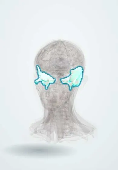
Representative model, for educational purposes only.