Intracerebral haemorrhage !
An intracerebral haemorrhage is bleeding inside the brain from a burst small artery; it causes sudden weakness, headache and drowsiness.
Updated 2026-10-29
Structures involved
Tap one to see what it is and open it in 3D.
In biochemistry
In the clinical exam
- Pronator drift
- Recognising a stroke: BE-FAST and the NIH Stroke Scale
- Intracranial hypertension syndrome
In physiology
- Motor cortex and the corticospinal tract
- Cerebral blood flow, brain metabolism and intracranial pressure
- Functions of the cerebellum
- The cerebral cortex and language
Why it happens
- Hemorrhage: petechiae, purpura, ecchymosis, hematoma, and what blood loss does
- Drugs of abuse: cocaine, opioids, amphetamines, cannabis
- Physiology of hypertension
- Blood vessels: artery, vein and capillary layers
What it is
Bleeding directly into the brain tissue from a ruptured small artery, causing a sudden focal deficit, headache and often reduced consciousness. It is a medical emergency with high mortality.
Who gets it
Accounts for a minority of strokes but a large share of stroke deaths. It is relatively more frequent in Latin American and other populations where hypertension is often undiagnosed or poorly controlled.
Causes
- Chronic hypertension (deep bleeds: basal ganglia, thalamus, pons, cerebellum)
- Cerebral amyloid angiopathy (lobar bleeds in older adults)
- Anticoagulant or antiplatelet use, clotting disorders
- Vascular malformations, tumours, haemorrhagic transformation of an infarct
- Stimulant drugs such as cocaine
Risk factors
- Hypertension
- Older age
- Anticoagulation
- Heavy alcohol use
- Smoking
- Previous intracerebral haemorrhage
Pathophysiology
Long-standing hypertension damages small penetrating arteries (lipohyalinosis, microaneurysms), which rupture. The haematoma compresses and injures brain, may keep growing in the first hours, and causes surrounding oedema. Large bleeds raise intracranial pressure and can extend into the ventricles, causing hydrocephalus.
Classification
- Deep (basal ganglia, especially putamen; thalamus)
- Lobar
- Cerebellar
- Brainstem (pons)
- With or without intraventricular extension
Symptoms and signs
- Sudden focal deficit (e.g. hemiparesis) that may worsen over minutes to hours
- Headache, nausea and vomiting
- Reduced level of consciousness
- Very high blood pressure on arrival
- Seizures in some lobar bleeds
Diagnosis
- Clinical
- Cannot be told apart from ischaemic stroke at the bedside; headache, vomiting and early drowsiness make it more likely. Severity is assessed with the Glasgow Coma Scale and scores such as the ICH score.
- Tests
- Blood count, clotting tests (INR), glucose, kidney function and toxicology when relevant.
- Imaging
- Non-contrast CT shows the bleed immediately. CT angiography looks for a spot sign or a vascular cause; MRI helps find amyloid angiopathy, tumours or cavernomas.
Differential diagnosis
- Ischaemic stroke
- Haemorrhagic transformation of an infarct
- Haemorrhagic tumour
- Traumatic contusion
- Subarachnoid haemorrhage
Goal of treatment
Stop the bleed from growing, protect the brain from secondary injury and raised pressure, and prevent recurrence.
Treatment
- Acute
- Airway protection if consciousness is reduced; careful, early blood pressure lowering as a core principle, with targets set by current guidelines.
- Medical
- Rapid reversal of anticoagulation with specific reversal agents or clotting factor concentrates.
- Follow-up
- Neurocritical or stroke unit care: neurological checks, glucose and temperature control, seizure treatment if they occur, thromboprophylaxis once the bleed is stable.
- Surgical
- Evacuation for selected cerebellar haemorrhages with deterioration or brainstem compression; external ventricular drain for hydrocephalus.
- Rehabilitation
- Early multidisciplinary rehabilitation.
Drugs for this condition
Complications
- Haematoma expansion
- Raised intracranial pressure and herniation
- Hydrocephalus
- Seizures
- Aspiration pneumonia and venous thromboembolism
Prognosis
Mortality is high, especially with large volume, deep or brainstem location, intraventricular blood and low Glasgow Coma Scale score. Survivors often have lasting disability, though some recover substantially.
Prevention
- Detection and control of hypertension
- Careful use of anticoagulants
- Limiting alcohol, avoiding stimulants
Test yourself
Think of the answer, then tap the question to see it.
What is the commonest cause of deep intracerebral haemorrhage?
Chronic hypertension.
Which site is most often involved in hypertensive haemorrhage?
The basal ganglia, especially the putamen; then thalamus, pons and cerebellum.
Which condition causes lobar haemorrhages in older adults?
Cerebral amyloid angiopathy.
Which haemorrhage location may need urgent surgical evacuation?
Cerebellar haemorrhage with deterioration or brainstem compression or hydrocephalus.
Related conditions
- Ischaemic stroke
- Subarachnoid haemorrhage
- Hypertensive heart disease
- Traumatic brain injury
- Arterial hypertension
- Epidural haematoma
Educational information, not medical advice. For any symptom, see a health professional.
Sources: Intracerebral Hemorrhagic Stroke (StatPearls (NCBI Bookshelf), 2026).
Spotted a mistake?We are always improving for the community. Tell us what is wrong and our team will review it.Report a mistake
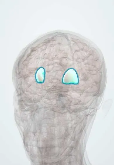
Representative model, for educational purposes only.