Conditions · Infectious and tropical diseases
Free-living amoeba infections (Naegleria meningoencephalitis, Acanthamoeba) !
Amoebas from water and soil: Naegleria enters the nose of swimmers and destroys the brain; Acanthamoeba infects the cornea of lens wearers.
Updated 2026-11-01
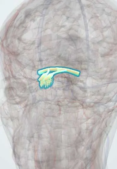
Structures involved
Tap one to see what it is and open it in 3D.
- Olfactory nerve (I)
- Cribriform plate
- Frontal lobe
- Cornea
- Nasal cavity
- Ethmoid bone
- Temporal lobe
- Dura mater
- White matter
Why it happens
- Patterns of necrosis: coagulative, liquefactive, caseous, fat, fibrinoid, gangrenous
- Cerebral blood flow, brain metabolism and intracranial pressure
- Olfactory epithelium and taste buds
- The eye wall: cornea, sclera, uvea and lens
What it is
Infections by amoebas that normally live in water and soil. Naegleria fowleri causes primary amoebic meningoencephalitis (PAM), a fulminant brain infection of healthy young people after freshwater exposure. Acanthamoeba causes a painful keratitis, mainly in contact lens wearers, and, like Balamuthia, a slow granulomatous amoebic encephalitis (GAE), usually in immunosuppressed people.
Who gets it
PAM is very rare but almost always fatal; the United States recorded 167 cases between 1962 and 2024 with four survivors, and cases are reported worldwide in hot climates, including Mexico, Venezuela and other parts of Latin America. Acanthamoeba keratitis is increasing with contact lens use and is the most frequent free-living amoeba infection. Balamuthia encephalitis is reported especially in Peru and other Latin American countries.
Causes
- Naegleria fowleri: thermophilic amoeba of warm fresh water (lakes, rivers, hot springs, poorly chlorinated pools)
- Acanthamoeba species: soil, dust, tap water, pools, hot tubs and lens cases
- Balamuthia mandrillaris: soil, causing skin lesions and encephalitis
- Naegleria enters by water forced into the nose; Acanthamoeba by the eye, skin wounds or lungs
Risk factors
- Naegleria: swimming, diving or water sports in warm fresh water in hot months, and nasal rinsing with untreated tap water
- Acanthamoeba keratitis: contact lens wear, rinsing lenses or cases with tap water, swimming or showering with lenses, and eye trauma
- GAE: HIV infection, transplantation, chemotherapy, diabetes and alcohol use disorder
- Children and young adults (PAM); not acquired by swallowing water or from other people
Pathophysiology
Naegleria trophozoites attach to the nasal mucosa, follow the olfactory nerve fibres through the cribriform plate and reach the olfactory bulbs and frontal lobes. There they feed on brain tissue and release cytolytic enzymes, causing haemorrhagic necrosis, intense neutrophilic inflammation, brain oedema and herniation within days. Acanthamoeba binds to damaged corneal epithelium, invades the stroma and the corneal nerves (causing severe pain) and forms resistant cysts. In GAE it spreads through the blood from the skin or lungs and causes a chronic granulomatous inflammation with necrotizing vasculitis.
Classification
- Primary amoebic meningoencephalitis by Naegleria fowleri (acute)
- Granulomatous amoebic encephalitis by Acanthamoeba or Balamuthia (subacute or chronic)
- Acanthamoeba keratitis
- Cutaneous and disseminated Acanthamoeba or Balamuthia infection
Symptoms and signs
- PAM: severe frontal headache, fever, nausea and vomiting 1 to 9 days after swimming
- PAM: stiff neck, altered smell or taste, confusion, seizures and rapid coma
- Keratitis: eye pain much stronger than the visible findings, redness, light sensitivity and blurred vision
- Keratitis: slow course over weeks, worsening despite usual antibacterial or antiviral drops
- GAE: headache, personality change, focal weakness and seizures over weeks to months
Diagnosis
- Clinical
- PAM looks like bacterial meningitis, so the clue is recent warm freshwater exposure; keratitis is suspected from lens use and pain out of proportion to the signs.
- Tests
- In PAM, cerebrospinal fluid is purulent, with high neutrophils and protein, low glucose and no bacteria; motile trophozoites are seen on a fresh wet mount of warm CSF, and PCR confirms the species. In keratitis, corneal scrapings or biopsy for stains, culture on non-nutrient agar with E. coli and PCR; in vivo confocal microscopy shows cysts. GAE needs brain biopsy or PCR.
- Imaging
- Brain CT or MRI in PAM shows oedema, basal meningeal enhancement and frontal haemorrhagic lesions; in GAE, multiple ring-enhancing or haemorrhagic lesions resembling abscesses or tumours.
Differential diagnosis
- Bacterial meningitis
- Herpes simplex and other viral encephalitis
- Toxoplasmosis, tuberculoma and cryptococcal meningitis (for GAE)
- Herpes simplex keratitis
- Bacterial and fungal keratitis
Goal of treatment
Start combination therapy as early as possible and control brain swelling in encephalitis; save vision and the cornea in keratitis.
Treatment
- Acute
- PAM is treated in intensive care with control of intracranial pressure (osmotic therapy, sedation, ventilation and sometimes induced hypothermia).
- Medical
- Combination therapy for PAM and GAE including miltefosine with amphotericin B, azoles, rifampin, azithromycin, pentamidine or flucytosine according to the organism, with advice from public health experts.
- Medical
- Acanthamoeba keratitis: frequent topical antiseptic drops (biguanides such as polyhexamethylene biguanide or chlorhexidine, sometimes with diamidines) for months; corneal transplant for scarring or perforation.
- Follow-up
- Neurological checks and intracranial pressure monitoring in encephalitis; frequent slit-lamp review in keratitis; report cases to public health to look for the water source.
Complications
- Brain oedema, herniation and death in PAM
- Permanent neurological damage in survivors
- Corneal scarring, perforation and loss of vision
- Need for corneal transplant or removal of the eye
- Dissemination to skin, sinuses and lungs in immunosuppressed people
Prognosis
PAM is fatal in over 95% of cases, usually within one to two weeks. GAE is also almost always fatal. Keratitis rarely threatens life but can cause permanent vision loss if diagnosis is delayed.
Prevention
- Avoiding diving into warm fresh water, holding the nose or using nose clips and keeping the head above water in hot springs
- Using boiled, filtered or distilled water for nasal rinses
- Never rinsing lenses or cases with tap water, removing lenses before swimming or showering and replacing cases regularly
- Adequate chlorination of pools
Test yourself
Think of the answer, then tap the question to see it.
How does Naegleria fowleri reach the brain?
Through the nose, along the olfactory nerve and across the cribriform plate to the olfactory bulbs and frontal lobes.
What CSF findings suggest primary amoebic meningoencephalitis?
Purulent CSF with neutrophils, low glucose and no bacteria, with motile amoebas on a fresh wet mount.
Who is at greatest risk of Acanthamoeba keratitis?
Contact lens wearers, especially those who expose lenses or cases to tap water or swim with lenses.
What is a classic clinical feature of Acanthamoeba keratitis?
Pain out of proportion to the findings and a ring-shaped corneal infiltrate.
Which drug is now included in combination therapy for free-living amoeba encephalitis?
Miltefosine.
Related conditions
- Bacterial meningitis
- Viral encephalitis (herpes simplex encephalitis)
- Keratitis and corneal ulcer
- Genital herpes (herpes simplex infection)
- Toxoplasmosis
- Cryptococcal meningitis
- HIV infection and AIDS
- Conjunctivitis
Educational information, not medical advice. For any symptom, see a health professional.
Sources: Naegleria fowleri Infections | Naegleria fowleri Infection | CDC (Centers for Disease Control and Prevention, 2024); Acanthamoeba Infections | Acanthamoeba Infections | CDC (Centers for Disease Control and Prevention, 2024).
Spotted a mistake?We are always improving for the community. Tell us what is wrong and our team will review it.Report a mistake
Representative model, for educational purposes only.