Conditions · Hematology and oncology
Hypereosinophilic syndrome !
Too many eosinophils, a type of white blood cell, build up in the blood and damage organs such as the heart, lungs, skin and nerves.
Updated 2026-11-01
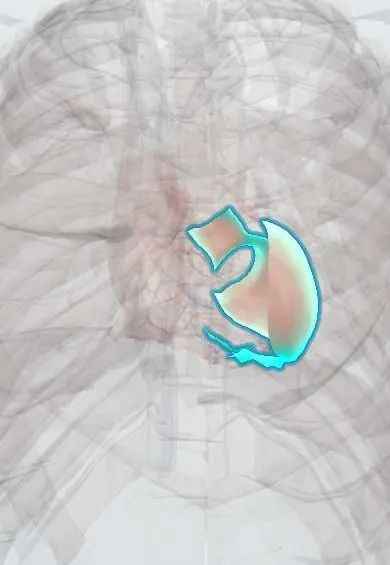
Structures involved
Tap one to see what it is and open it in 3D.
- Left ventricle
- Right ventricle
- Mitral posterior leaflet
- Tricuspid septal leaflet
- Left ventricle papillary muscles
Why it happens
- When leukocytes injure tissue: proteases, oxidants and NETs
- Fibrosis in organs: TGF-β and the myofibroblast
- White blood cells: types and functions
What it is
A group of disorders defined by persistent marked blood eosinophilia (at least 1.5 × 10⁹/L on two occasions, or striking tissue eosinophilia) with organ damage caused by the eosinophils, after other causes such as parasites, allergy and drugs have been excluded or identified. Heart involvement (endomyocardial fibrosis, Löffler endocarditis) is the main cause of death.
Who gets it
Rare, with an estimated prevalence of under 1 to a few per 100,000. Usually diagnosed between 20 and 50 years; the myeloproliferative FIP1L1-PDGFRA form occurs almost only in men. In Colombia and the tropics, helminth infections (Strongyloides, Toxocara, hookworm) are far more common causes of eosinophilia and must be ruled out first.
Causes
- Primary (neoplastic): clonal eosinophils from a myeloid neoplasm, classically the FIP1L1-PDGFRA fusion on chromosome 4q12; also PDGFRB, FGFR1 or JAK2 rearrangements and chronic eosinophilic leukaemia
- Lymphocytic variant: abnormal T cells that secrete interleukin-5
- Secondary (reactive): parasites, drug reactions, allergic disease, vasculitis and some cancers
- Idiopathic: no cause found after full evaluation
Risk factors
- Male sex (myeloproliferative form)
- Age 20–50
- For secondary eosinophilia: travel or residence in areas with helminth infection, new medications
Pathophysiology
Eosinophils release toxic granule proteins (major basic protein, eosinophil cationic protein), reactive oxygen species and cytokines that injure tissue. In the heart, damage follows three stages: acute necrosis of the endocardium and myocardium, mural thrombus formation in the ventricles, and late fibrosis that stiffens the ventricles (restrictive cardiomyopathy) and tethers the mitral and tricuspid valve leaflets, causing regurgitation. Eosinophils also promote clotting, so arterial and venous thrombosis and cardioembolic strokes are common.
Classification
- Myeloid/primary HES, including FIP1L1-PDGFRA-positive disease
- Lymphocytic-variant HES
- Secondary (reactive) hypereosinophilia
- Familial HES
- Idiopathic HES
- Organ-restricted forms, such as eosinophilic pneumonia or eosinophilic oesophagitis, are related but separate
Symptoms and signs
- Fatigue, fever, cough and muscle aches
- Itchy rash, urticaria or angio-oedema
- Breathlessness and wheeze from lung infiltrates
- Heart failure symptoms: breathlessness, oedema, palpitations
- Numbness or weakness from peripheral neuropathy; confusion or stroke-like episodes
- Diarrhoea and abdominal pain from gut infiltration
- Enlarged spleen in the myeloproliferative form
Diagnosis
- Clinical
- Suspect it in persistent eosinophilia with organ involvement. First search for common reactive causes: parasites, drugs and allergic disease. Then assess every organ at risk, especially the heart.
- Tests
- Full blood count with smear repeated over time; stool examination and Strongyloides serology; troponin, ECG; vitamin B12 and tryptase (raised in myeloid forms); FIP1L1-PDGFRA testing in blood by FISH or PCR; bone marrow biopsy with cytogenetics; T-cell receptor clonality and flow cytometry for the lymphocytic variant; tissue biopsy of affected organs.
- Imaging
- Echocardiography to look for apical thrombus, endomyocardial thickening and valve regurgitation; cardiac MRI with late gadolinium enhancement detects early endomyocardial fibrosis; chest CT for lung infiltrates.
Differential diagnosis
- Parasitic infection (Strongyloides, Toxocara, hookworm, Fasciola)
- Drug reaction with eosinophilia and systemic symptoms (DRESS)
- Eosinophilic granulomatosis with polyangiitis
- Eosinophilic pneumonia
- Systemic mastocytosis and chronic myeloid leukaemia
- Hodgkin lymphoma and T-cell lymphoma
Goal of treatment
Lower the eosinophil count quickly to stop organ damage, especially to the heart, and treat the underlying cause or clone.
Treatment
- Medical
- FIP1L1-PDGFRA-positive and other PDGFR-rearranged disease: imatinib, a tyrosine kinase inhibitor, usually produces complete remission.
- Acute
- Most other forms, and emergencies with organ damage: high-dose systemic corticosteroids, after or together with empirical ivermectin when Strongyloides cannot be excluded.
- Medical
- Steroid-sparing therapy: anti-interleukin-5 monoclonal antibodies (such as mepolizumab), hydroxyurea or interferon alfa; chemotherapy or stem cell transplantation for aggressive refractory disease.
- Medical
- Organ support: heart failure treatment, anticoagulation for intracardiac thrombus, and valve repair or replacement for severe fibrotic regurgitation.
- Follow-up
- Serial eosinophil counts, troponin and echocardiography to detect cardiac involvement early.
Complications
- Endomyocardial fibrosis with restrictive cardiomyopathy and heart failure
- Mitral and tricuspid regurgitation
- Ventricular thrombus, stroke and other emboli; venous thrombosis
- Pulmonary fibrosis
- Transformation to acute leukaemia in myeloid forms
- Strongyloides hyperinfection after corticosteroids
Prognosis
Prognosis has improved greatly: most patients with FIP1L1-PDGFRA disease remain in remission on imatinib, and many others are controlled with corticosteroids and biologics. Cardiac involvement and myeloid forms without a targetable fusion carry the worst outcome.
Test yourself
Think of the answer, then tap the question to see it.
What eosinophil count defines hypereosinophilia in hypereosinophilic syndrome?
At least 1.5 × 10⁹/L on two occasions, with eosinophil-mediated organ damage.
Which fusion gene identifies the imatinib-responsive form of HES?
FIP1L1-PDGFRA.
What cardiac lesion is typical of hypereosinophilic syndrome?
Endomyocardial fibrosis (Löffler endocarditis) with ventricular thrombus and restrictive physiology.
Which parasite must be excluded or treated before giving corticosteroids for eosinophilia?
Strongyloides stercoralis, because steroids can trigger hyperinfection.
Related conditions
- Eosinophilic pneumonias (including Löffler syndrome)
- Restrictive cardiomyopathy (amyloid, sarcoid, haemochromatosis)
- Eosinophilic granulomatosis with polyangiitis (Churg–Strauss)
- Strongyloidiasis
- Toxocariasis (visceral and ocular larva migrans)
- Mastocytosis (cutaneous and systemic)
- Chronic myeloid leukaemia
- Heart failure
- Mitral regurgitation
- Ischaemic stroke
- Deep vein thrombosis
- Peripheral neuropathy (diabetic and other)
Educational information, not medical advice. For any symptom, see a health professional.
Sources: Hypereosinophilic Syndrome (National Organization for Rare Disorders (NORD), 2026).
Spotted a mistake?We are always improving for the community. Tell us what is wrong and our team will review it.Report a mistake
Representative model, for educational purposes only.