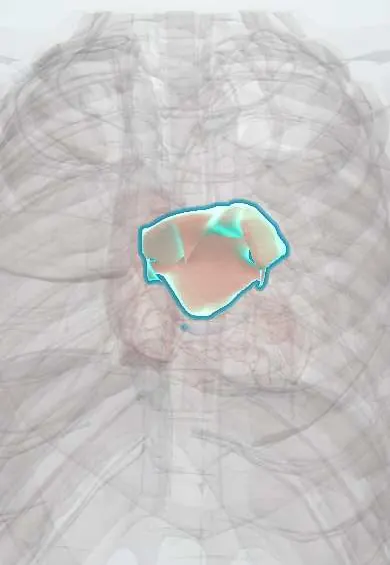
Restrictive cardiomyopathy (amyloid, sarcoid, haemochromatosis) !
Stiff heart muscle that fills poorly although it squeezes almost normally, causing heart failure. Amyloid is the most common cause.
Updated 2026-11-01
Structures involved
Tap one to see what it is and open it in 3D.
- Left atrium
- Right atrium
- Left ventricle
- Right ventricle
- Interventricular septum
- Pericardium
- Atrioventricular (AV) node
- Inferior vena cava
In the clinical exam
Why it happens
- Fibrosis in organs: TGF-β and the myofibroblast
- Granulomatous inflammation: caseating vs non-caseating, the causes to know
- The ventricular pressure–volume loop
- Transthyretin (prealbumin)
- Iron metabolism
What it is
Restrictive cardiomyopathy is a disease of the heart muscle in which the ventricles are abnormally stiff, so diastolic filling is impaired, while ventricular size and systolic function are normal or near normal early on. Filling pressures rise, the atria enlarge greatly, and heart failure with preserved ejection fraction follows. It is the least common of the main cardiomyopathies.
Who gets it
Rare overall, though cardiac amyloidosis (especially wild-type transthyretin amyloid in older men) is increasingly recognised with modern imaging. Endomyocardial fibrosis is a cause in tropical regions of Africa, Asia and South America. Specific data for Colombia are limited; hereditary transthyretin amyloidosis variants have been described in Latin America.
Causes
- Infiltrative: amyloidosis (light-chain AL, wild-type or hereditary transthyretin ATTR), sarcoidosis
- Storage: hereditary haemochromatosis, Fabry disease, glycogen storage diseases
- Endomyocardial: endomyocardial fibrosis, hypereosinophilic syndrome (Löffler endocarditis), carcinoid heart disease
- Other: idiopathic or familial, mediastinal radiation, anthracyclines, systemic sclerosis
Risk factors
- Older age and male sex (wild-type ATTR amyloid)
- Plasma cell dyscrasia or multiple myeloma (AL amyloid)
- Family history of amyloidosis or haemochromatosis
- Chest radiation therapy
- Living in tropical regions (endomyocardial fibrosis)
Pathophysiology
Infiltration (amyloid, granulomas), deposition (iron) or fibrosis of the myocardium or endocardium makes the ventricles non-compliant. A small rise in volume causes a steep rise in pressure, so filling occurs mostly in early diastole and then stops abruptly. High pressures back up into both atria and the pulmonary and systemic veins. Stroke volume is fixed and low, so cardiac output depends on heart rate; atrial fibrillation and conduction disease are common because the infiltration also affects the atria and the conduction system.
Classification
- Infiltrative (amyloid, sarcoid)
- Storage diseases (haemochromatosis, Fabry, glycogen storage)
- Endomyocardial (endomyocardial fibrosis, hypereosinophilic syndrome, carcinoid, radiation)
- Idiopathic or familial
Symptoms and signs
- Exertional dyspnoea, fatigue and poor exercise tolerance
- Predominantly right-sided congestion: leg oedema, ascites, hepatomegaly, raised jugular venous pressure
- Orthopnoea and paroxysmal nocturnal dyspnoea
- Palpitations from atrial fibrillation; syncope from conduction block or arrhythmia
- Clues to amyloid: bilateral carpal tunnel syndrome, macroglossia, periorbital purpura, neuropathy, intolerance to antihypertensives
Diagnosis
- Clinical
- Suspect it in heart failure with preserved ejection fraction, a non-dilated heart with very large atria and marked right-sided congestion. Raised jugular pressure that rises on inspiration (Kussmaul sign) and an S3 or S4 may be present.
- Tests
- ECG: low QRS voltage despite thick walls (classic for amyloid), pseudo-infarct pattern, AV block, atrial fibrillation. BNP or NT-proBNP and troponin raised. Serum and urine free light chains and immunofixation for AL amyloid; ferritin and transferrin saturation for haemochromatosis; genetic testing for hereditary ATTR. Endomyocardial biopsy when the cause remains uncertain. Cardiac catheterisation shows a «dip and plateau» (square root) ventricular pressure, as in constriction; concordant respiratory changes in right and left ventricular systolic pressures (discordant in constriction) help tell them apart.
- Imaging
- Echocardiography: normal-sized ventricles with thick walls (in infiltration), biatrial enlargement, restrictive filling pattern and markedly reduced tissue Doppler velocities; apical sparing of longitudinal strain in amyloid. Cardiac MRI with late gadolinium enhancement (diffuse subendocardial in amyloid, patchy in sarcoid) and T2* for iron. Bone scintigraphy (technetium pyrophosphate) diagnoses ATTR amyloid without biopsy when light chains are normal.
Differential diagnosis
- Constrictive pericarditis
- Hypertrophic cardiomyopathy
- Hypertensive heart disease with diastolic dysfunction
- Cor pulmonale and pulmonary hypertension
- Cirrhosis or nephrotic syndrome with ascites and oedema
Goal of treatment
Relieve congestion without lowering preload too much, treat the specific cause, control rhythm and conduction problems, and prevent thromboembolism.
Treatment
- Medical
- Careful diuretics for congestion; patients are preload dependent, so over-diuresis causes hypotension and kidney injury. Vasodilators and rate-slowing drugs are often poorly tolerated.
- Medical
- Anticoagulation for atrial fibrillation, and in amyloid often regardless of the CHA2DS2-VASc score because atrial thrombus is common.
- Medical
- Cause-specific therapy: transthyretin stabilisers or silencers for ATTR amyloid; plasma cell–directed chemotherapy for AL amyloid; immunosuppression with corticosteroids for cardiac sarcoidosis; phlebotomy or iron chelation for haemochromatosis; enzyme replacement for Fabry disease.
- Procedure
- Permanent pacemaker for symptomatic bradycardia or AV block; implantable defibrillator in selected patients (especially sarcoidosis).
- Surgical
- Heart transplantation for advanced disease in selected patients; endocardial resection in endomyocardial fibrosis.
- Follow-up
- Daily weight, fluid balance, blood pressure (orthostatic hypotension is common in amyloid), kidney function and electrolytes; rhythm monitoring for AV block and atrial fibrillation.
Complications
- Progressive heart failure and low cardiac output
- Atrial fibrillation and thromboembolism (stroke)
- AV block, ventricular arrhythmias and sudden death
- Cardiac cirrhosis and protein-losing congestion
- Kidney injury from low output or over-diuresis
Prognosis
Generally poor once heart failure develops, especially in AL amyloid. Outcomes are better when a treatable cause is found early: ATTR therapies slow progression, haemochromatosis can improve with iron removal, and sarcoidosis may respond to immunosuppression.
Prevention
- Early screening of relatives in hereditary haemochromatosis and hereditary ATTR amyloid
- Early recognition of red-flag signs (carpal tunnel syndrome, low ECG voltage with thick walls) to treat before advanced disease
- Limiting cardiac radiation exposure in cancer therapy
Test yourself
Think of the answer, then tap the question to see it.
What is the main haemodynamic problem in restrictive cardiomyopathy?
Impaired diastolic filling of stiff ventricles with normal or near-normal systolic function.
Which ECG finding with thick ventricular walls on echo suggests cardiac amyloidosis?
Low QRS voltage.
Which test can diagnose transthyretin cardiac amyloid without a biopsy?
Bone (technetium pyrophosphate) scintigraphy, once light-chain amyloid is excluded.
Which condition is the main differential diagnosis and may need surgery instead?
Constrictive pericarditis.
Why must diuretics be used carefully?
The stiff ventricles are preload dependent, so excessive diuresis lowers cardiac output and causes hypotension.
Related conditions
- Amyloidosis
- Hereditary hemochromatosis
- Sarcoidosis
- Constrictive pericarditis
- Heart failure
- Atrial fibrillation
- Atrioventricular (heart) block
- Hypertrophic cardiomyopathy
- Dilated cardiomyopathy
- Systemic sclerosis (scleroderma, including CREST)
Educational information, not medical advice. For any symptom, see a health professional.
Sources: Restrictive cardiomyopathy (MedlinePlus, US National Library of Medicine, 2026).
Spotted a mistake?We are always improving for the community. Tell us what is wrong and our team will review it.Report a mistake
Representative model, for educational purposes only.