Hypertensive emergency and urgency !
A hypertensive crisis is a very high blood pressure. It is an emergency when it is acutely damaging the brain, heart, kidneys or vessels.
Updated 2026-11-01
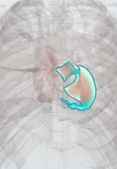
Structures involved
Tap one to see what it is and open it in 3D.
In the clinical exam
- Hypertensive emergency: target-organ signs
- Blood pressure measurement
- Fundoscopy: the optic disc, papilledema and retinopathy
Why it happens
- Patterns of necrosis: coagulative, liquefactive, caseous, fat, fibrinoid, gangrenous
- Physiology of hypertension
- Local and humoral control of blood flow
- Cerebral blood flow, brain metabolism and intracranial pressure
- Blood vessels: artery, vein and capillary layers
What it is
Severely raised blood pressure, commonly above about 180/120 mmHg. It is a hypertensive emergency when there is new or worsening target-organ damage, and a hypertensive urgency (severe asymptomatic hypertension) when there is none.
Who gets it
Arterial hypertension is very common in Colombia and Latin America and is often undiagnosed or poorly controlled, so hypertensive crises are frequent in emergency departments. Most are urgencies; true emergencies are fewer but carry high mortality.
Causes
- Poorly controlled chronic hypertension or stopping antihypertensive drugs (the most common cause)
- Kidney disease: acute glomerulonephritis, renal artery stenosis
- Pre-eclampsia and eclampsia
- Catecholamine excess: pheochromocytoma, cocaine or amphetamine use, abrupt clonidine withdrawal
- Acute pain, anxiety or urinary retention (raise blood pressure without true emergency)
Risk factors
- Untreated or poorly adherent hypertension
- Chronic kidney disease
- Pregnancy
- Stimulant drug use
- Limited access to follow-up and medicines
Pathophysiology
A sudden rise in vascular resistance overwhelms autoregulation in arterioles. Endothelial injury, fibrinoid necrosis and microthrombi cause ischaemia in the brain, retina, kidney and heart, and pressure-driven natriuresis causes volume depletion that activates the renin-angiotensin system, driving a further rise in pressure. Chronic hypertension shifts autoregulation upward, so a too-fast fall in pressure can also cause ischaemia.
Classification
- Hypertensive emergency: severe hypertension with acute target-organ damage
- Hypertensive urgency: severe hypertension without acute target-organ damage
- Malignant hypertension: severe hypertension with retinal haemorrhages, exudates or papilloedema
Symptoms and signs
- Severe headache, blurred vision, confusion or seizures (hypertensive encephalopathy)
- Chest pain or tearing pain radiating to the back
- Dyspnoea and orthopnoea from acute pulmonary oedema
- Focal neurological deficit
- Reduced urine output or blood in the urine
- Often no symptoms in a hypertensive urgency
Diagnosis
- Clinical
- Repeated blood pressure in both arms with a proper cuff, and a focused search for target-organ damage: neurological examination, fundoscopy (haemorrhages, exudates, papilloedema), heart and lung examination, pulses and urine output.
- Tests
- ECG and troponin; creatinine, electrolytes and urinalysis (protein, red cells); complete blood count and smear for haemolysis; pregnancy test in women of childbearing age; urine toxicology when stimulants are suspected.
- Imaging
- Chest X-ray for pulmonary oedema or a widened mediastinum; brain CT or MRI with neurological signs; CT angiography or echocardiography when aortic dissection is suspected.
Differential diagnosis
- Pain- or anxiety-driven rise in blood pressure
- Primary stroke or intracerebral haemorrhage with reactive hypertension
- Aortic dissection
- Pre-eclampsia
- Stimulant intoxication or alcohol withdrawal
Goal of treatment
In an emergency, lower blood pressure in a controlled, gradual way (usually by no more than about a quarter in the first hour) to stop organ damage without causing ischaemia; in an urgency, lower it over hours to days with oral treatment and secure follow-up.
Treatment
- Acute
- Hypertensive emergency: admission to intensive care with an arterial line and short-acting, titratable intravenous agents (for example labetalol, nicardipine, clevidipine, esmolol or nitroprusside), chosen by the organ involved.
- Acute
- Special situations: aortic dissection needs rapid lowering of heart rate and pressure with a beta-blocker first; acute ischaemic stroke usually allows higher pressures; pulmonary oedema benefits from nitrates and loop diuretics; pre-eclampsia uses magnesium sulfate plus labetalol or hydralazine.
- Acute
- Catecholamine excess (pheochromocytoma, cocaine): alpha-blockade (phentolamine) and benzodiazepines; a beta-blocker alone is avoided.
- Medical
- Hypertensive urgency: restart or adjust oral antihypertensives and lower pressure gradually, avoiding rapid-acting agents that cause sudden drops.
- Follow-up
- Frequent blood pressure and neurological checks, urine output, kidney function and cardiac monitoring; then education on adherence and close outpatient follow-up.
Complications
- Ischaemic stroke, intracerebral haemorrhage and hypertensive encephalopathy
- Acute coronary syndrome and acute heart failure with pulmonary oedema
- Aortic dissection
- Acute kidney injury
- Eclampsia and HELLP syndrome in pregnancy
- Organ ischaemia from lowering blood pressure too quickly
Prognosis
Untreated hypertensive emergency has high short-term mortality; with timely treatment most survive, but long-term cardiovascular and kidney risk stays high. Urgencies have a good short-term outlook when follow-up is secured.
Prevention
- Diagnosis and sustained control of chronic hypertension
- Adherence to antihypertensive treatment and regular follow-up
- Avoiding stimulant drugs
Test yourself
Think of the answer, then tap the question to see it.
What distinguishes a hypertensive emergency from a hypertensive urgency?
Acute target-organ damage (brain, heart, aorta, kidney, retina) is present in an emergency and absent in an urgency.
How fast is blood pressure usually lowered in a hypertensive emergency?
Gradually: by no more than about 25% in the first hour, with exceptions such as aortic dissection, which needs faster lowering.
Why is a beta-blocker alone avoided in cocaine- or pheochromocytoma-induced crisis?
Unopposed alpha-adrenergic vasoconstriction can raise blood pressure further; alpha-blockade and benzodiazepines are used instead.
Which fundoscopic findings define malignant hypertension?
Retinal haemorrhages, cotton-wool spots or exudates, and papilloedema.
Related conditions
- Arterial hypertension
- Aortic dissection
- Ischaemic stroke
- Intracerebral haemorrhage
- Preeclampsia
- Phaeochromocytoma
- Acute kidney injury
- Heart failure
- Myocardial Infarction (Heart Attack)
- Hypertensive heart disease
Educational information, not medical advice. For any symptom, see a health professional.
Sources: Malignant hypertension: MedlinePlus Medical Encyclopedia (MedlinePlus, US National Library of Medicine, 2026).
Spotted a mistake?We are always improving for the community. Tell us what is wrong and our team will review it.Report a mistake
Representative model, for educational purposes only.