
Inferior vena cava filters
A small metal trap placed in the body's main vein to catch leg clots before they reach the lungs, used when blood thinners cannot be given.
What it is
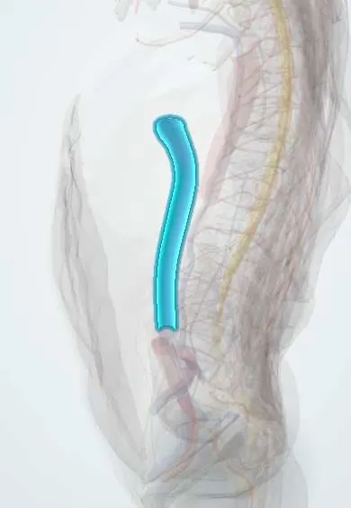
A wire device placed through a vein, usually in the inferior vena cava just below the renal veins, to stop large clots from the legs or pelvis travelling to the lungs. It is a substitute for anticoagulation when anticoagulation cannot be given, not an addition to it. Most filters used today are retrievable.
Who gets it
Filter use spread widely after retrievable designs arrived, despite no proof that filters lower mortality. After a US FDA warning in 2010 about complications of filters left in too long, guidelines narrowed the indications and use fell. Retrieval rates have been low in many series (a mean of about one third in one analysis), mainly because no follow-up was planned.
Causes
- Classic (agreed) indication: acute proximal deep vein thrombosis or pulmonary embolism with a contraindication to anticoagulation, such as active bleeding, recent intracranial haemorrhage, very low platelets or a high-bleeding-risk procedure
- Failure or complication of anticoagulation: recurrent embolism or clot extension despite adequate anticoagulation, or major bleeding on treatment
- Debated (expanded) indications: massive or high-risk embolism with residual leg clot, free-floating iliofemoral or caval thrombus, very poor cardiopulmonary reserve
- Prophylactic use without clot (major trauma, bariatric or orthopaedic surgery): not supported by trials; guidelines differ or advise against it
Risk factors
- Long dwell time (the main driver of thrombosis, fracture, migration and failed retrieval)
- No planned follow-up or retrieval date
- Filter tilt or a hook embedded in the caval wall
- Ongoing clot risk without anticoagulation
Pathophysiology
The filter's struts sit against the caval wall and trap emboli coming up from the leg and pelvic veins. It does not dissolve clot or treat the cause. Blood flowing past a foreign body and trapped clot can thrombose the cava, and venous collaterals can develop that let emboli bypass a long-standing filter. Over time the struts become covered with tissue and can tilt, erode through the wall into nearby organs, or break; this incorporation is why late retrieval is harder.
Classification
- Permanent filters: meant to stay for life; generally not removable through a catheter
- Retrievable (optional) filters: have a hook for snaring and can be removed, or left if still needed
- Convertible filters: open into a non-filtering shape when protection is no longer needed
- Temporary filters: attached to a catheter or the skin and removed with it
- By position: infrarenal (standard); suprarenal for thrombus in the infrarenal cava or renal or gonadal veins, pregnancy or caval anomalies
Symptoms and signs
- Usually none after placement
- Bilateral leg swelling or a heavy, congested lower body (caval thrombosis)
- Abdominal or back pain (strut perforation into nearby organs)
- Chest pain, palpitations or breathlessness (fragment migration to the heart or lungs)
- Soreness at the groin or neck access site
Diagnosis
- Clinical
- Confirm the venous thromboembolism and that anticoagulation is truly contraindicated (or has failed) before placement. Ask the retrieval question at the same time: when will the contraindication end, who will remove the filter and when.
- Tests
- Blood count and coagulation tests to define the bleeding contraindication; review of platelet count and anticoagulation history in suspected failure; kidney function before contrast.
- Imaging
- Compression ultrasound and CT pulmonary angiography confirm the clot. Placement is under fluoroscopy or ultrasound through the femoral or jugular vein, with a cavogram to define the renal veins and caval size. Afterwards, plain films or CT show tilt, fracture, migration or perforation; duplex or CT venography shows caval thrombosis.
Differential diagnosis
- Anticoagulation alone (the standard when it can be given)
- Catheter-directed or systemic thrombolysis for high-risk pulmonary embolism
- Surgical or catheter embolectomy
- Mechanical prophylaxis (intermittent pneumatic compression) when the problem is prevention, not established clot
Who should get a filter? The strength of each indication
| Indication | Example | Evidence and consensus | Usual answer |
|---|---|---|---|
| Clot plus a contraindication to anticoagulation | Proximal DVT or PE with active bleeding or recent brain haemorrhage | Agreed by all major societies; no randomised trial (it would be unethical) | Yes: retrievable filter, anticoagulate once safe, then remove |
| Anticoagulation failed or caused harm | New PE or clot extension despite adequate treatment; major bleeding on treatment | Broad consensus | Usually yes, after checking the anticoagulation was really adequate |
| Added to working anticoagulation | High-risk PE with residual leg clot, free-floating iliac clot | PREPIC trials: fewer early emboli, no lasting or survival benefit; PREPIC2 showed no benefit | Generally no; individual decision only |
| Prophylaxis without clot | Major trauma, bariatric surgery, cancer | Trials and reviews show no clear benefit and more DVT | No in most guidelines |
Goal of treatment
Prevent pulmonary embolism while anticoagulation is impossible, then restore anticoagulation and remove the filter as soon as the danger has passed.
Treatment
- Procedure
- Retrievable filter placed through the femoral or internal jugular vein, usually just below the renal veins; suprarenal only for specific reasons (thrombus below or at the renal veins, pregnancy, caval anomaly).
- Procedure
- Permanent filter only when the contraindication to anticoagulation is expected to be lifelong and retrieval is unlikely, for example with limited life expectancy.
- Medical
- Start or restart anticoagulation as soon as the contraindication resolves: a filter does not treat the clot, and anticoagulation is still needed for the deep vein thrombosis itself.
- Follow-up
- Plan retrieval at placement: record the filter, tell the patient it is temporary, book review, and remove it once anticoagulation is running or the risk has passed. Retrieval is more successful when attempted within about 3 months; experts suggest about 90–120 days. The lack of planned follow-up is the main reason filters stay in.
- Procedure
- Difficult retrieval of tilted or embedded filters: advanced endovascular techniques (loop snare, forceps, laser sheath) in experienced centres, and open or laparoscopic removal when these fail.
Complications
- Caval thrombosis and filter occlusion, with leg swelling and, rarely, severe venous congestion and kidney injury
- Recurrent deep vein thrombosis (more frequent with a filter in long-term trial follow-up)
- Migration of the filter or a fragment, occasionally to the heart or lungs
- Strut fracture and embolisation of fragments
- Perforation of the caval wall, usually silent, sometimes into the duodenum, aorta or vertebra
- Access-site bleeding, haematoma or arteriovenous fistula, and misplacement during insertion
- Failed retrieval when the filter is tilted, embedded or full of clot
Common traps
- A filter treats the deep vein thrombosis: it only catches emboli; the clot is treated with anticoagulation, which is started as soon as it is safe
- Adding a filter to anticoagulation gives extra protection: in PREPIC it cut early emboli but not survival, and raised later DVT; PREPIC2 found no benefit from retrievable filters
- A retrievable filter can be left because it is retrievable: retrieval gets harder and complications rise with dwell time; remove it once it is no longer needed
- A filter prevents all pulmonary emboli: emboli can pass through collaterals or a clotted filter, and new clot can form on the filter itself
- High-risk trauma or bariatric patients need a prophylactic filter: trials have not shown a clear benefit, and most guidelines advise against routine use
Prognosis
Prognosis is mainly that of the underlying clot and the reason anticoagulation was impossible. Short dwell times and timely retrieval keep filter-related harm low; long-dwelling filters accumulate thrombosis, perforation and fracture.
Prevention
- Place filters only for clear indications, mainly clot with a contraindication to anticoagulation
- Choose a retrievable filter when the contraindication is temporary
- Set a retrieval plan and date at the time of placement
- Resume anticoagulation as soon as it is safe
Test yourself
Think of the answer, then tap the question to see it.
What is the main agreed indication for an inferior vena cava filter?
Acute proximal DVT or pulmonary embolism in a patient who cannot be anticoagulated (for example active bleeding or recent brain haemorrhage).
Why is anticoagulation restarted after a filter is placed once it is safe?
A filter only catches emboli. It does not treat the clot or stop new clots, and the filter itself can thrombose.
What did the PREPIC trials show about adding a filter to anticoagulation?
PREPIC: fewer early emboli but no survival benefit and more DVT later. PREPIC2 (retrievable filters): no benefit. So filters are not added routinely.
Why should a retrievable filter be removed early rather than late?
Thrombosis, fracture, migration and perforation rise with dwell time, and the filter becomes embedded, so retrieval succeeds less often.
When is a filter placed above the renal veins?
When thrombus reaches the infrarenal cava or renal or gonadal veins, in pregnancy, or with a caval anomaly such as a duplicated cava.
Connections
Educational information, not medical advice. For any symptom, see a health professional.
Sources: Inferior Vena Cava Filters: An Overview (Methodist DeBakey Cardiovascular Journal, 2024); Inferior Vena Cava Filters: A Clinical Review and Future Perspectives (Journal of Clinical Medicine, 2024); Pulmonary Embolism (PE) (MSD Manual Professional Edition, 2025).
Spotted a mistake?We are always improving for the community. Tell us what is wrong and our team will review it.Report a mistake
Representative model, for educational purposes only.