
Mirizzi syndrome
A gallstone stuck in the gallbladder neck presses on the main bile duct next to it and causes jaundice. It makes gallbladder surgery risky.
What it is
A rare complication of gallstones in which a stone impacted in the gallbladder neck (Hartmann's pouch) or cystic duct compresses the common hepatic duct, causing obstructive jaundice. Long-standing pressure and inflammation can erode the wall and create a cholecystobiliary fistula, or a fistula into the bowel.
Who gets it
Found in about 0.05–6% of patients with gallstones; about 0.5% of cholecystectomies in Europe and about 5% in Asia and Central and South America, and about 1% of ERCPs. More common in women and older adults, often after long-standing gallstone disease.
Causes
- A large gallstone impacted in Hartmann's pouch or the cystic duct
- Anatomy that brings the ducts close: a long cystic duct running parallel to the common hepatic duct with a low insertion, or a short cystic duct
- Chronic inflammation from repeated cholecystitis
- Pressure necrosis of the duct wall, leading to a cholecystobiliary or cholecystoenteric fistula
Risk factors
- Long-standing gallstones
- Repeated attacks of cholecystitis
- Cystic duct variants (long, parallel, low insertion)
- Female sex and older age
Pathophysiology
Stone impacted in the gallbladder neck or cystic duct → gallbladder inflammation and extrinsic pressure on the adjacent common hepatic duct → bile backs up into the liver (obstructive jaundice, dilated ducts above, normal common bile duct below) → persistent pressure and inflammation cause necrosis and ulceration of the duct wall → a cholecystobiliary fistula forms and widens (Csendes II to IV) → the gallbladder and bile duct fuse into one inflamed mass; a fistula can also open into the duodenum or colon, and a stone that passes into the bowel can cause gallstone ileus.
Classification
- Csendes type I: external compression of the common hepatic duct by a stone, no fistula
- Type II: cholecystobiliary fistula involving less than one third of the duct circumference
- Type III: fistula involving up to two thirds of the circumference
- Type IV: fistula with complete destruction of the bile duct wall
- Type V: any of the above plus a cholecystoenteric fistula, Va without and Vb with gallstone ileus
- Older McSherry classification: type I compression, type II fistula
Symptoms and signs
- Right upper quadrant pain, often recurrent (most patients)
- Obstructive jaundice (about half to most patients)
- Nausea and vomiting
- Fever and chills when cholangitis develops
- Dark urine, pale stools, itching
- Positive Murphy sign; sometimes a hard gallbladder mass that feels like cancer
Diagnosis
- Clinical
- Suspect it when a patient with gallstones or cholecystitis is jaundiced but no stone is seen in the common bile duct. Only about a third to two thirds of cases are recognised before surgery; the rest are found during the operation.
- Tests
- Cholestatic liver tests: raised alkaline phosphatase, GGT and bilirubin, with milder aminotransferase rises; white cell count often raised. CA 19-9 can be raised by the obstruction itself and does not separate Mirizzi syndrome from cancer. At surgery, frozen section of the gallbladder wall is used to exclude a coexisting carcinoma.
- Imaging
- Ultrasound first: a stone in the gallbladder neck with dilated intrahepatic ducts and common hepatic duct above it and a normal-calibre common bile duct below, but it often misses the diagnosis. MRCP shows the narrowing and the dilated ducts above it well and is the preferred non-invasive test. CT mainly helps exclude a tumour. ERCP is the reference and can also treat (stent, stone removal), and cholangioscopy can break the stone.
Differential diagnosis
- Choledocholithiasis (stone inside the common bile duct)
- Gallbladder cancer
- Cholangiocarcinoma
- Pancreatic head cancer
- Acute cholangitis from other causes
- Primary sclerosing cholangitis
Gallstones and jaundice: where is the problem?
| Feature | Mirizzi syndrome | Choledocholithiasis | Gallbladder cancer |
|---|---|---|---|
| Where the stone or lesion is | Stone in the gallbladder neck or cystic duct, pressing from outside | Stone inside the common bile duct | Tumour of the gallbladder wall, invading the ducts or liver |
| Ducts on imaging | Dilated above the common hepatic duct, normal common bile duct below | Common bile duct dilated down to the stone | Mass at the gallbladder; ducts dilated above it |
| Course | Recurrent pain and cholecystitis, then jaundice | Colicky pain, jaundice that fluctuates as the stone moves | Often silent, then painless jaundice and weight loss |
| Best test | MRCP, ERCP | MRCP, endoscopic ultrasound, ERCP | CT and MRI staging; histology |
| Treatment | Cholecystectomy, often subtotal or open; bile duct repair or bypass for big fistulas | ERCP stone removal, then cholecystectomy | Radical resection if possible |
Goal of treatment
Remove the gallbladder and the impacted stone, repair or bypass the damaged duct according to its type, and avoid a bile duct injury; relieve jaundice and treat cholangitis first when present.
Treatment
- Procedure
- ERCP before surgery for jaundice or cholangitis: sphincterotomy, stone removal and a stent keep the duct open and make the operation safer; cholangioscopy-guided lithotripsy can break the impacted stone in selected cases.
- Surgical
- Type I: cholecystectomy, laparoscopic in experienced hands, with a low threshold for converting to open surgery or doing a subtotal cholecystectomy when the anatomy is unclear.
- Surgical
- Types II and III: subtotal cholecystectomy, using a cuff of gallbladder wall to close the fistula (choledochoplasty); a Roux-en-Y hepaticojejunostomy for large defects.
- Surgical
- Type IV: cholecystectomy with Roux-en-Y hepaticojejunostomy. Type V: open surgery to close the bowel fistula, and removal of the stone from the bowel in gallstone ileus.
- Follow-up
- After surgery, watch for bile leak, cholangitis and late bile duct stricture. Nursing care: drain output and colour, jaundice, fever and liver tests.
Complications
- Bile duct injury during cholecystectomy (much more likely when the syndrome is not recognised beforehand)
- Acute cholangitis
- Cholecystobiliary and cholecystoenteric fistula
- Gallstone ileus
- Secondary biliary cirrhosis from long-standing obstruction
- Coexisting gallbladder cancer
Common traps
- Jaundice with gallstones means a stone in the common bile duct: a stone in the gallbladder neck can squeeze the common hepatic duct from outside while the common bile duct is empty
- Mirizzi syndrome is just a hard cholecystectomy: dense inflammation hides the hepatocystic triangle and the duct may be part of the gallbladder wall, so bile duct injury is common; subtotal cholecystectomy or open surgery is the safe bail-out
- A raised CA 19-9 settles that it is cancer: biliary obstruction itself raises CA 19-9; the two can look the same, so the gallbladder wall is checked by frozen section
- Normal ultrasound bile ducts exclude it: ultrasound misses many cases; MRCP or ERCP shows the narrowing
- Once cancer is excluded it is a benign problem with no cancer link: gallbladder cancer is found more often in Mirizzi syndrome than in simple gallstone disease, especially in older patients with long histories
Prognosis
Good after surgery when the type is recognised and the duct is repaired or bypassed, with low mortality in experienced centres. Unrecognised disease raises the risk of bile duct injury, conversion to open surgery and later stricture. A coexisting gallbladder cancer sets the prognosis when present.
Prevention
- Timely cholecystectomy for symptomatic gallstones before stones and inflammation have time to erode the ducts
- Preoperative MRCP or ERCP when a gallstone patient is jaundiced, so the surgeon can plan the operation
Test yourself
Think of the answer, then tap the question to see it.
In Mirizzi syndrome, why is the patient jaundiced although the common bile duct contains no stone?
A stone impacted in the gallbladder neck or cystic duct compresses the adjacent common hepatic duct from outside.
What does MRCP typically show in Mirizzi syndrome?
A stone in the gallbladder neck, a narrowed common hepatic duct with dilated ducts above it, and a normal-calibre common bile duct below.
What separates Csendes type I from types II to IV?
Type I is compression only; types II to IV have a cholecystobiliary fistula that destroys a third, two thirds or all of the duct wall.
Why is cholecystectomy in Mirizzi syndrome dangerous, and what is the usual bail-out?
Inflammation and fusion hide the anatomy, so bile duct injury is common; subtotal cholecystectomy or conversion to open surgery is safer.
Which cancer can Mirizzi syndrome mimic or coexist with, and why is CA 19-9 unhelpful?
Gallbladder cancer (also cholangiocarcinoma); CA 19-9 rises with biliary obstruction alone, so it cannot separate them.
Connections
Educational information, not medical advice. For any symptom, see a health professional.
Sources: The Current Approach to the Diagnosis and Classification of Mirizzi Syndrome (Diagnostics (Basel), 2021); Mirizzi Syndrome-The Past, Present, and Future (Medicina (Kaunas), 2023); Current trends in the management of Mirizzi Syndrome: A review of literature (Medicine (Baltimore), 2018).
Spotted a mistake?We are always improving for the community. Tell us what is wrong and our team will review it.Report a mistake
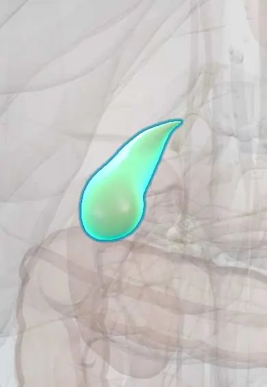
Representative model, for educational purposes only.