Conditions · Infectious and tropical diseases
Nontuberculous mycobacterial infections (MAC, M. abscessus, Buruli ulcer) !
Mycobacteria other than tuberculosis, found in water and soil, can infect damaged lungs, lymph nodes, skin and, in advanced HIV, the whole body.
Updated 2026-11-01
Structures involved
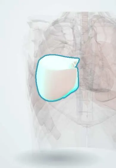
Tap one to see what it is and open it in 3D.
- Right middle lobe
- Neck lymph nodes
- Left upper lobe
- Segmental bronchi
- Lobar bronchi
- Mediastinal lymph nodes
- Skin
Why it happens
- Granulomatous inflammation: caseating vs non-caseating, the causes to know
- Special stains in diagnosis: Congo red, Prussian blue, PAS, Ziehl–Neelsen, Gram
- Airway defenses, cough and mucociliary clearance
- Innate and adaptive immunity: overview
What it is
Infections by environmental mycobacteria other than the tuberculosis and leprosy bacilli. They cause slowly progressive lung disease, neck lymphadenitis in children, skin and soft tissue infections after procedures or injuries, and disseminated disease in severe immunosuppression. Mycobacterium avium complex (MAC), M. abscessus and M. ulcerans (Buruli ulcer) are the main exam examples.
Who gets it
NTM are found in soil, tap water, showers and biofilms worldwide, and lung disease is increasing, especially in older adults with structural lung disease. In Colombia and other Latin American countries, outbreaks of skin infection by rapidly growing mycobacteria such as M. abscessus have followed cosmetic procedures (liposuction, mesotherapy, injections) done with poorly sterilised equipment. Buruli ulcer occurs mainly in West and Central Africa, with smaller foci elsewhere, including South America.
Causes
- Slow growers: M. avium complex, M. kansasii, M. ulcerans
- Rapid growers: M. abscessus, M. chelonae, M. fortuitum
- Acquired from the environment (water aerosols, soil, contaminated medical or cosmetic equipment), not usually from other people
- More than 190 species described
Risk factors
- Bronchiectasis, COPD, cystic fibrosis, previous tuberculosis
- Thin, older women with nodular bronchiectasis (classic MAC lung disease)
- Advanced HIV with a very low CD4 count (disseminated MAC)
- Cosmetic surgery, injections, tattoos or catheters with contaminated water or equipment
- Immunosuppressive drugs, including anti-TNF therapy
- Defects in the interferon-gamma / IL-12 pathway
Pathophysiology
NTM are inhaled or inoculated and taken up by macrophages. Control depends on Th1 immunity (IL-12, interferon-gamma). In damaged airways they colonise and cause chronic granulomatous inflammation, nodules, bronchiectasis and cavities. With very low CD4 counts, MAC spreads through the blood to marrow, liver, spleen and nodes. M. ulcerans produces a toxin (mycolactone) that destroys tissue and suppresses immunity locally, so its large ulcers are painless.
Classification
- Pulmonary: fibrocavitary (upper lobe, smokers, like tuberculosis) or nodular-bronchiectatic (middle lobe and lingula, older women)
- Lymphadenitis: unilateral neck node in young children
- Skin and soft tissue: post-procedure abscesses, Buruli ulcer, fish tank granuloma (M. marinum)
- Disseminated: advanced HIV and other severe immunosuppression
Symptoms and signs
- Chronic cough, sputum, fatigue, weight loss and sometimes haemoptysis in lung disease
- Painless, firm, unilateral neck node in a well child that may turn violaceous and drain
- Tender nodules, abscesses and draining sinuses weeks after a cosmetic procedure or injection
- Painless nodule or swelling on a limb that becomes a large ulcer with undermined edges (Buruli ulcer)
- Fever, night sweats, diarrhoea, anaemia and hepatosplenomegaly in disseminated MAC
Diagnosis
- Clinical
- Lung disease needs compatible symptoms, imaging and microbiology together, after excluding tuberculosis; a single positive sputum culture may be contamination. Skin infection is suspected when abscesses fail standard antibiotics after a procedure.
- Tests
- Acid-fast smear and mycobacterial culture with species identification (at least two positive sputum cultures, or one bronchoscopic sample, for lung disease); susceptibility testing, including macrolide resistance; blood culture for mycobacteria in disseminated disease; tissue biopsy and culture for skin and nodes; PCR (IS2404) for Buruli ulcer; tuberculosis tests to exclude M. tuberculosis.
- Imaging
- Chest CT: tree-in-bud nodules and bronchiectasis in the middle lobe and lingula, or upper lobe cavities; ultrasound or CT of skin collections and nodes.
Differential diagnosis
- Pulmonary tuberculosis
- Bronchiectasis from other causes, aspergillosis, lung cancer
- Tuberculous lymphadenitis (scrofula) or cat-scratch disease in neck nodes
- Bacterial skin abscess, sporotrichosis, cutaneous leishmaniasis
- Disseminated histoplasmosis or lymphoma in advanced HIV
Goal of treatment
Treat only clinically significant disease, with long multidrug regimens guided by species and susceptibility; remove infected tissue or devices; and restore immunity in people with HIV.
Treatment
- Medical
- MAC lung disease: a macrolide (azithromycin or clarithromycin) plus ethambutol and a rifamycin, continued for about a year after sputum becomes negative; amikacin is added for cavitary or refractory disease.
- Medical
- M. abscessus: induction with intravenous drugs (amikacin plus a carbapenem or cefoxitin, or tigecycline) and oral agents chosen by susceptibility; disseminated MAC: macrolide plus ethambutol with antiretroviral therapy; Buruli ulcer: rifampicin plus clarithromycin for 8 weeks.
- Surgical
- Complete excision of NTM lymphadenitis in children; debridement of skin abscesses and removal of infected implants or catheters; lung resection for localised refractory disease; reconstructive surgery for Buruli ulcer.
- Follow-up
- Monthly sputum cultures; hearing, vision and liver tests during long therapy; wound care and physiotherapy to prevent contractures in Buruli ulcer.
Drugs for this condition
Complications
- Progressive lung destruction and respiratory failure
- Macrolide-resistant disease that is hard to cure
- Disfiguring scars and draining sinuses after cosmetic-procedure infections
- Contractures and disability from Buruli ulcer
- Immune reconstitution inflammatory syndrome after starting antiretroviral therapy
Prognosis
Treatment is long and side effects are common. Nodular MAC disease often responds, but relapse and reinfection are frequent. M. abscessus lung disease has the lowest cure rates. Lymphadenitis in children is usually cured by excision, and small Buruli lesions heal well with antibiotics.
Prevention
- Strict sterilisation and safe water use in surgical and cosmetic procedures
- Antiretroviral therapy for people with HIV
- Early treatment of Buruli lesions to prevent disability
Test yourself
Think of the answer, then tap the question to see it.
Which patient has classic nodular-bronchiectatic MAC lung disease?
A thin, older woman with bronchiectasis in the right middle lobe and lingula.
When does disseminated MAC typically occur?
In advanced HIV with a very low CD4 count.
Which mycobacterium causes outbreaks after cosmetic procedures?
Rapid growers such as Mycobacterium abscessus.
What is the treatment of NTM neck lymphadenitis in a child?
Complete surgical excision of the node.
Why is the Buruli ulcer painless?
Mycobacterium ulcerans produces mycolactone, a toxin that destroys tissue and blocks pain and local immunity.
Related conditions
- Tuberculosis
- Bronchiectasis
- HIV infection and AIDS
- Chronic obstructive pulmonary disease (COPD)
- Cystic fibrosis
- Leprosy (Hansen disease)
- Leishmaniasis
- Abscess, furuncle and carbuncle
- Histoplasmosis
Educational information, not medical advice. For any symptom, see a health professional.
Sources: About Nontuberculous Mycobacteria (NTM) Infections | NTM | CDC (Centers for Disease Control and Prevention, 2024); Buruli ulcer (Mycobacterium ulcerans infection) (World Health Organization, 2025).
Spotted a mistake?We are always improving for the community. Tell us what is wrong and our team will review it.Report a mistake
Representative model, for educational purposes only.