Conditions · Hematology and oncology
Paroxysmal nocturnal haemoglobinuria !
A rare acquired blood disease in which the body's own complement system destroys red cells, causing dark urine, anaemia and dangerous blood clots.
Updated 2026-11-01
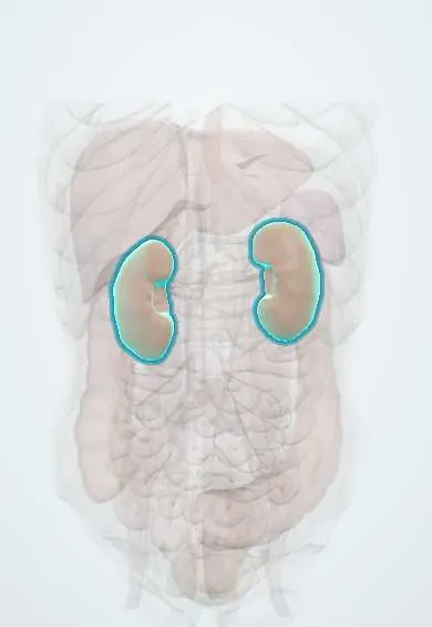
Structures involved
Tap one to see what it is and open it in 3D.
In immunology
Why it happens
What it is
An acquired clonal disorder of blood stem cells in which a PIGA gene mutation leaves red cells, white cells and platelets without GPI-anchored proteins (including CD55 and CD59) that normally protect them from complement. It causes intravascular haemolysis, thrombosis and bone marrow failure.
Who gets it
Very rare (a few cases per million people per year), affecting men and women equally, mostly young adults in their 30s. It is closely linked to aplastic anaemia: a small PNH clone is found in many patients with aplastic anaemia. In Latin America, as elsewhere, it is often diagnosed late because haemoglobinuria is not always present and flow cytometry is available mainly in referral centres.
Causes
- Acquired somatic mutation of the X-linked PIGA gene in a haematopoietic stem cell (not inherited)
- Clonal expansion of PNH cells, often in a marrow damaged by immune attack (aplastic anaemia)
- Triggers of haemolytic crises: infections, surgery, pregnancy, strenuous exercise and transfusion
Risk factors
- Aplastic anaemia
- Myelodysplastic syndrome (less often)
- Young adult age
- Pregnancy and infections (precipitate crises and thrombosis)
Pathophysiology
Without CD55 (decay-accelerating factor) and CD59 (membrane inhibitor of reactive lysis), red cells cannot stop the complement cascade on their surface, and the membrane attack complex punches holes in them. Haemolysis occurs inside vessels, releasing free haemoglobin that is lost in the urine (haemoglobinuria, haemosiderinuria) and scavenges nitric oxide, causing smooth muscle spasm: abdominal pain, difficulty swallowing, erectile dysfunction and pulmonary hypertension. Activated platelets, free haemoglobin and microparticles make the blood highly prone to clot, especially in unusual veins. The defective stem cells also contribute to marrow failure.
Classification
- Classic (haemolytic) PNH: marked haemolysis with a cellular marrow
- PNH in the setting of another marrow failure disorder (aplastic anaemia or myelodysplastic syndrome)
- Subclinical PNH: a small clone without clinical haemolysis
Symptoms and signs
- Dark, cola-coloured urine, classically in the morning
- Tiredness, pallor and breathlessness from anaemia
- Jaundice
- Episodes of abdominal pain and difficulty swallowing
- Erectile dysfunction
- Bruising, bleeding or infections when the marrow fails
Diagnosis
- Clinical
- Suspect PNH in Coombs-negative haemolytic anaemia, haemolysis with thrombosis in unusual sites, haemolysis with cytopenias, or any patient with aplastic anaemia.
- Tests
- Flow cytometry of peripheral blood is the diagnostic test: it shows red cells and granulocytes lacking CD55 and CD59, or fluorescent aerolysin (FLAER) binding loss, and measures clone size. Haemolysis markers: high LDH (often very high), high reticulocytes, low haptoglobin and high indirect bilirubin, with a negative direct antiglobulin (Coombs) test. Urine shows haemoglobin and haemosiderin. Iron deficiency is common from urinary iron loss. Bone marrow examination looks for associated aplastic anaemia or myelodysplasia.
- Imaging
- Doppler ultrasound, CT or MRI when thrombosis is suspected (hepatic, portal, mesenteric and cerebral veins); echocardiography for pulmonary hypertension.
Differential diagnosis
- Autoimmune haemolytic anaemia (Coombs positive)
- G6PD deficiency and other inherited haemolytic anaemias
- Thrombotic microangiopathies (TTP, haemolytic uraemic syndrome)
- Aplastic anaemia or myelodysplastic syndrome without a PNH clone
- Other causes of dark urine: myoglobinuria, haematuria
Goal of treatment
Stop complement-mediated haemolysis, prevent and treat thrombosis, support the blood count and, in selected patients, cure the disease with stem cell transplantation.
Treatment
- Medical
- Terminal complement (C5) inhibitors such as eculizumab or ravulizumab block intravascular haemolysis and markedly reduce thrombosis; proximal complement inhibitors (C3 or factor B/D inhibitors) treat persistent extravascular haemolysis.
- Medical
- Meningococcal vaccination before starting complement inhibitors, often with antibiotic prophylaxis, because these drugs greatly increase the risk of Neisseria infection.
- Medical
- Anticoagulation for established thrombosis; folic acid and iron supplementation; red cell transfusions for symptomatic anaemia.
- Procedure
- Allogeneic haematopoietic stem cell transplantation, the only cure, mainly for patients with severe marrow failure or refractory disease; immunosuppressive therapy when PNH coexists with aplastic anaemia.
- Follow-up
- Nursing priorities: urine colour, signs of thrombosis (abdominal pain, headache, leg swelling), haemoglobin and LDH trends, renal function, and fever on complement inhibitors treated as an emergency.
Complications
- Venous thrombosis in unusual sites (hepatic, portal, mesenteric, cerebral veins), the main cause of death
- Chronic kidney disease and acute kidney injury from haemoglobin deposition
- Pulmonary hypertension
- Bone marrow failure (aplastic anaemia) and evolution to myelodysplastic syndrome or acute leukaemia
- Meningococcal infection on complement inhibitor therapy
Prognosis
Before complement inhibitors, median survival was roughly 10 to 20 years, and thrombosis was the main cause of death. With C5 inhibitor therapy survival approaches that of the general population in many patients, although some still need transfusions because of extravascular haemolysis or marrow failure.
Test yourself
Think of the answer, then tap the question to see it.
Which gene is mutated in paroxysmal nocturnal haemoglobinuria?
PIGA, an X-linked gene needed to make GPI anchors, through an acquired somatic mutation in a stem cell.
Which two protective surface proteins are missing on PNH red cells?
CD55 (decay-accelerating factor) and CD59 (membrane inhibitor of reactive lysis).
What is the diagnostic test of choice for PNH?
Flow cytometry of peripheral blood showing loss of CD55/CD59 or FLAER binding.
What is the leading cause of death in untreated PNH?
Thrombosis, especially of the hepatic veins (Budd-Chiari syndrome) and other abdominal or cerebral veins.
Which vaccination is required before starting a complement C5 inhibitor?
Meningococcal vaccination, because complement blockade greatly raises the risk of Neisseria meningitidis infection.
Related conditions
- Aplastic anaemia
- Myelodysplastic syndromes
- Autoimmune haemolytic anaemia
- Budd-Chiari syndrome
- Portal vein thrombosis
- Cerebral venous sinus thrombosis
- Deep vein thrombosis
- Pulmonary embolism
- Chronic kidney disease
- Iron deficiency anaemia
- Bacterial meningitis
Educational information, not medical advice. For any symptom, see a health professional.
Sources: Paroxysmal nocturnal hemoglobinuria (PNH) (MedlinePlus (U.S. National Library of Medicine), 2026); Paroxysmal nocturnal hemoglobinuria (MedlinePlus Genetics (U.S. National Library of Medicine), 2022).
Spotted a mistake?We are always improving for the community. Tell us what is wrong and our team will review it.Report a mistake
Representative model, for educational purposes only.