
Pseudoxanthoma elasticum
An inherited disease in which calcium builds up in elastic tissue. It shows as yellow bumps on the neck and can damage the eyes and arteries.
Quick facts
- Key signs
- Small yellowish · Lax · Calf pain on walking
- Diagnosis
- Genetic testing for ABCC6 variants
Red flags ↓
No doses: the clinician sets them by the current guideline.
What it is
An autosomal recessive disorder caused by ABCC6 variants in which calcium and other minerals deposit in elastic fibres of the skin, Bruch's membrane of the eye and the walls of medium-sized arteries. It causes yellow papules on the neck and flexures, angioid streaks with vision loss, and early arterial disease.
Who gets it
Rare: estimates range from about one in 25 000 to one in 56 000 people worldwide, with founder clusters such as the Afrikaner population of South Africa. It is diagnosed about twice as often in women, perhaps because the skin changes are noticed more. Skin lesions usually appear in childhood or adolescence, and eye complications commonly start in adult life.
Causes
- Biallelic pathogenic variants in ABCC6 (chromosome 16), encoding a transporter expressed mainly in the liver and kidney; more than 300 variants are known
- Autosomal recessive inheritance; pseudodominant transmission occurs when a carrier has children with an affected partner
- A PXE-like picture also occurs with ENPP1 or GGCX variants and in some haemoglobinopathies such as beta-thalassaemia and sickle cell disease
Risk factors
- Affected sibling or parents who are carriers (consanguinity raises the chance)
- For complications: smoking, hypertension, raised cholesterol and diabetes (arteries); blows to the head and eye (retinal haemorrhage)
Pathophysiology
ABCC6 deficiency in liver cells → less ATP released into the blood → less of it converted to pyrophosphate → low plasma pyrophosphate, the main circulating inhibitor of soft-tissue mineralisation → calcium phosphate deposits on elastic fibres, which fragment → skin: calcified dermal elastic fibres produce yellowish papules and lax redundant skin in areas of movement (neck, axillae, groin, flexures) → eye: Bruch's membrane calcifies, first as peau d'orange mottling, then cracks into angioid streaks radiating from the optic disc; new vessels from the choroid grow through the cracks (choroidal neovascularisation), bleed and scar, destroying central vision → arteries: calcification of the internal elastic lamina of medium-sized arteries narrows them, causing claudication (legs most affected), and less often coronary disease, stroke and bleeding from fragile gastrointestinal vessels. Disturbed lipid handling adds to early atherosclerosis.
Classification
- Skin: papules and plaques, lax redundant skin, rarely perforating lesions
- Eye: peau d'orange, angioid streaks, comet-tail lesions, choroidal neovascularisation and macular scarring
- Vascular: peripheral artery disease, coronary disease, stroke, hypertension
- Gastrointestinal: occasional bleeding from calcified vessels of the digestive tract
- Diagnostic certainty: definite (skin histology plus eye findings, or two pathogenic ABCC6 variants) versus probable
Symptoms and signs
- Small yellowish or skin-coloured papules on the side of the neck, armpits, groins and elbow and knee creases, coalescing into larger plaques
- Lax, wrinkled, redundant skin in the same places with time
- Yellowish papules inside the lower lip
- Distorted or blurred central vision, a central blind spot, reduced night vision
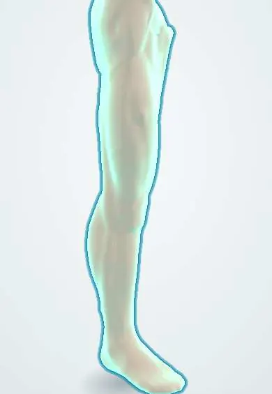
- Calf pain on walking (intermittent claudication)
- Vomiting blood or black stools from gastrointestinal bleeding (uncommon)
Diagnosis
- Clinical
- Look for the characteristic flexural papules, examine the inside of the lower lip, and dilate the pupils to look for peau d'orange and angioid streaks. Ask about vision, claudication, gastrointestinal bleeding and family history; examine pulses and blood pressure.
- Tests
- Skin biopsy from a lesion (or a flexural site) shows fragmented, calcified elastic fibres in the dermis, with calcium shown by the von Kossa stain. Genetic testing for ABCC6 variants confirms and allows family testing. Lipids, glucose and kidney function for cardiovascular risk.
- Imaging
- Ophthalmology with fundus photography, autofluorescence, optical coherence tomography and angiography to find choroidal neovascularisation. Arterial Doppler ultrasound or CT for peripheral artery calcification; endoscopy for gastrointestinal bleeding.
Differential diagnosis
- PXE-like disorders: ENPP1 or GGCX deficiency, beta-thalassaemia and sickle cell disease (also cause angioid streaks)
- Solar elastosis and other acquired yellowish papules on sun-exposed skin
- Cutis laxa (lax skin without calcification or eye disease)
- Ehlers–Danlos syndrome and Marfan syndrome (other elastic and connective tissue disorders)
- Age-related macular degeneration (choroidal neovascularisation in older people, without angioid streaks)
Angioid streaks or yellow neck papules: which disease?
| Condition | Skin | Eye | Other clues | Confirmation |
|---|---|---|---|---|
| Pseudoxanthoma elasticum | Yellow papules and lax skin on neck and flexures | Peau d'orange, angioid streaks, choroidal neovascularisation | Claudication, gastrointestinal bleeding, family history | Skin biopsy with calcium stain; ABCC6 testing |
| Sickle cell disease or beta-thalassaemia | Usually normal | Angioid streaks can occur | Anaemia, haemolysis, crises | Haemoglobin electrophoresis |
| Ehlers–Danlos syndrome | Hyperextensible, fragile skin, poor scars | Usually no angioid streaks | Joint hypermobility | Clinical criteria and genetic testing |
| Age-related macular degeneration | Normal | Drusen and choroidal neovascularisation in older age, no streaks | Age over 50, smoking | Retinal imaging |
Goal of treatment
There is no cure: protect vision by finding and treating choroidal neovascularisation early, slow arterial disease by controlling cardiovascular risk, and manage bleeding and skin concerns.
Treatment
- Procedure
- Intravitreal anti-VEGF injections, the first-choice treatment for choroidal neovascularisation secondary to angioid streaks, started at the first sign of fluid or new vessels and repeated as needed; older laser and photodynamic treatments are largely outdated.
- Lifestyle
- Not smoking, regular walking exercise for claudication, a healthy diet and weight; avoiding contact sports and wearing protective eyewear, because minor eye trauma can tear Bruch's membrane and bleed; daily self-testing of central vision with an Amsler grid.
- Medical
- Treat hypertension, lipids (statins) and diabetes as in established cardiovascular disease. Antiplatelet or anticoagulant drugs only when clearly indicated and after blood pressure is controlled, because of retinal bleeding risk. Restricting dietary calcium has not been shown to help, and etidronate remains investigational.
- Surgical
- Cosmetic surgery for redundant neck skin in selected patients (wound healing can be poor); endoscopic treatment of gastrointestinal bleeding; revascularisation rarely needed because collaterals usually compensate.
- Follow-up
- Regular eye examinations with retinal imaging, yearly cardiovascular review, and genetic counselling for the family.
Complications
- Loss of central vision from choroidal neovascularisation, haemorrhage and scarring
- Peripheral artery disease with claudication
- Coronary artery disease, myocardial infarction and stroke at a young age
- Gastrointestinal bleeding
- Hypertension
- Cosmetic concern from lax skin
Common traps
- PXE is a skin disease: the defect is in a liver transporter that sets plasma pyrophosphate, and the skin is only the visible marker of a body-wide calcification of elastic fibres
- Angioid streaks themselves cause the vision loss: the streaks are cracks in Bruch's membrane; vision is lost when new vessels grow through them, bleed and scar, which anti-VEGF injections can prevent
- Angioid streaks always mean PXE: they also occur in sickle cell disease, beta-thalassaemia and Paget disease of bone
- A low-calcium diet will stop the calcification: dietary calcium restriction has not been shown to help; the problem is the low pyrophosphate guard
- Yellow neck papules in a young woman are only cosmetic: they are a warning sign that calls for an eye examination and a cardiovascular check
Prognosis
Life expectancy is usually near normal, but quality of life is often reduced. The main disability is loss of central vision, which anti-VEGF treatment started early can largely preserve. Arterial disease mostly causes claudication; serious coronary and cerebrovascular events are less common.
Prevention
- Genetic counselling and testing of siblings
- Early, regular eye examination and Amsler grid self-testing
- Control of cardiovascular risk factors from a young age
- Avoiding head and eye trauma
Test yourself
Think of the answer, then tap the question to see it.
Where is the ABCC6 transporter mainly expressed, and why does that matter in PXE?
In the liver; its loss lowers plasma pyrophosphate, so calcification of elastic fibres happens in distant tissues (skin, eye, arteries).
What eye finding links PXE with sickle cell disease and Paget disease of bone?
Angioid streaks: cracks in a calcified Bruch's membrane radiating from the optic disc.
What is the main cause of vision loss in PXE and how is it treated?
Choroidal neovascularisation through angioid streaks, with bleeding and scarring; it is treated with intravitreal anti-VEGF injections.
Which biopsy finding confirms PXE?
Fragmented, calcified elastic fibres in the dermis, with the calcium shown by the von Kossa stain.
Which arterial territory is usually most affected in PXE, and what symptom does it cause?
The arteries of the legs; calcification and narrowing cause intermittent claudication.
Connections
Educational information, not medical advice. For any symptom, see a health professional.
Sources: Pseudoxanthoma elasticum (MedlinePlus Genetics, US National Library of Medicine, 2026); Therapy of pseudoxanthoma elasticum: current knowledge and future perspectives (Biomedicines) (Europe PMC, 2021); Angioid streaks remain a challenge in diagnosis, management, and treatment (Vision) (Europe PMC, 2024); ABCC6 and pseudoxanthoma elasticum: the face of a rare disease from genetics to advocacy (International Journal of Molecular Sciences) (Europe PMC, 2017).
Spotted a mistake?We are always improving for the community. Tell us what is wrong and our team will review it.Report a mistake
Representative model, for educational purposes only.