Conditions · Surgery and trauma
Renal trauma !
Injury to a kidney from a blow, fall or wound. Blood in the urine is the key sign; most stable patients heal without surgery.
Updated 2026-11-01
Structures involved
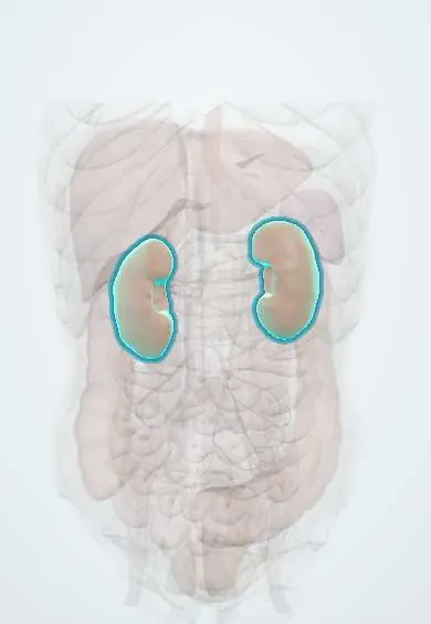
Tap one to see what it is and open it in 3D.
- Kidneys
- Left renal artery
- Right renal artery
- Left renal vein
- Right renal vein
- Renal pelvis
- Ureters
- Left adrenal gland
- Spleen
- Liver
Why it happens
- Hemorrhage: petechiae, purpura, ecchymosis, hematoma, and what blood loss does
- Infarction: red vs white, and what decides the damage
What it is
Injury to the kidney parenchyma, collecting system or renal vessels from blunt, penetrating or deceleration trauma. The kidney is the most commonly injured organ of the urinary tract; most injuries are low grade and are managed without surgery.
Who gets it
Kidney and urinary tract injuries appear in roughly one in ten to one in five abdominal trauma cases. Blunt mechanisms predominate worldwide, but in Colombia penetrating injuries from stab and gunshot wounds make up an important share in urban trauma centres. Children are relatively more vulnerable because their kidneys are less protected.
Causes
- Blunt trauma: road traffic crashes, falls, direct blows to the flank or back
- Rapid deceleration stretching the renal pedicle (arterial intimal tear, thrombosis)
- Penetrating trauma: stab and gunshot wounds of the flank, back or abdomen
- Sports injuries and iatrogenic injury (biopsy, percutaneous procedures)
Risk factors
- Fractures of the lower ribs or lumbar transverse processes
- Childhood (less perirenal fat and rib protection)
- Pre-existing kidney abnormalities: hydronephrosis, tumours, horseshoe kidney
- High-energy mechanism or multiple trauma
Pathophysiology
Direct impact crushes the kidney against the ribs and spine, causing contusion or laceration. Deceleration stretches the fixed renal artery, tearing its intima and causing thrombosis and loss of perfusion of the kidney. Deep lacerations into the collecting system leak urine (urinoma), and an ischaemic kidney can trigger the renin-angiotensin system and cause hypertension later.
Classification
- AAST grade I: contusion or non-expanding subcapsular haematoma
- Grade II: laceration under 1 cm without urine leak
- Grade III: laceration over 1 cm without collecting system injury
- Grade IV: laceration into the collecting system, or segmental vascular injury
- Grade V: shattered kidney or avulsion of the renal hilum
- WSES classes I–IV add haemodynamic status to the anatomic grade
Symptoms and signs
- Haematuria, visible or microscopic (may be absent in pedicle injuries)
- Flank or abdominal pain
- Flank bruising or abrasion
- Palpable flank mass (haematoma)
- Signs of blood loss
Diagnosis
- Clinical
- Suspect it with flank trauma, lower rib fractures or a deceleration mechanism. Imaging is indicated in stable patients with visible haematuria, with microscopic haematuria plus hypotension, with a suggestive mechanism, and in all penetrating flank or back wounds.
- Tests
- Urinalysis for blood, serial haemoglobin, blood group and crossmatch, creatinine and electrolytes.
- Imaging
- Contrast-enhanced CT with delayed (excretory) images is the test of choice in stable patients: it grades the injury, shows urine leaks and checks the other kidney. Angiography for active bleeding. Unstable patients go to surgery, where intraoperative one-shot urography may be used.
Differential diagnosis
- Adrenal haemorrhage
- Splenic or liver injury
- Ureteric or bladder injury as the source of haematuria
- Lumbar spine or rib fracture pain
- Pre-existing kidney stone or tumour bleeding
Goal of treatment
Control bleeding, preserve as much kidney function as possible, drain any urine leak, and prevent late complications such as hypertension.
Treatment
- Conservative
- Non-operative management for haemodynamically stable patients: bed rest at first, serial haemoglobin and examination, and monitoring of urine; this is the standard for most grades.
- Procedure
- Selective angioembolisation for active contrast extravasation, pseudoaneurysm or arteriovenous fistula, or for ongoing bleeding during non-operative management.
- Procedure
- Ureteral stent or percutaneous drainage for persistent urine leak or symptomatic urinoma.
- Surgical
- Operative exploration for haemodynamic instability, expanding or pulsatile haematoma or renal pedicle avulsion: renorrhaphy, partial nephrectomy or nephrectomy.
- Follow-up
- Nursing priorities: vital signs, urine output and colour, serial haemoglobin, flank pain and fever; blood pressure follow-up after high-grade injuries.
Complications
- Delayed bleeding
- Urinoma and perirenal abscess
- Arteriovenous fistula or pseudoaneurysm
- Renovascular (renin-mediated) hypertension
- Loss of renal function or of the kidney
Prognosis
Low-grade injuries heal well and kidney preservation is achieved in most patients managed non-operatively. Grade V injuries, pedicle injuries and instability carry the highest risk of nephrectomy; long-term blood pressure checks are advised after high-grade injury.
Prevention
- Road safety and seatbelt use
- Motorcycle protective gear
- Violence prevention
Test yourself
Think of the answer, then tap the question to see it.
What is the imaging test of choice for a stable patient with suspected renal trauma?
Contrast-enhanced CT with delayed excretory images to grade the injury and detect urine leak.
Does the absence of haematuria exclude a serious renal injury?
No. Renal artery thrombosis or pedicle avulsion can present without haematuria.
What is the standard management of a stable patient with renal trauma?
Non-operative management with monitoring, adding angioembolisation for active bleeding when needed.
Which late complication links an ischaemic kidney with raised blood pressure?
Renovascular hypertension from activation of the renin-angiotensin system.
Related conditions
- Blunt abdominal trauma
- Penetrating trauma (stab and gunshot wounds)
- Rib fractures
- Arterial hypertension
- Acute kidney injury
- Bladder and urethral injury
- Splenic injury
- Liver injury
- Major trauma (polytrauma): ATLS primary survey
Educational information, not medical advice. For any symptom, see a health professional.
Sources: Kidney and uro-trauma: WSES-AAST guidelines (World Journal of Emergency Surgery, 2019); Urotrauma Guideline - American Urological Association (American Urological Association, 2020).
Spotted a mistake?We are always improving for the community. Tell us what is wrong and our team will review it.Report a mistake
Representative model, for educational purposes only.