Conditions · Surgery and trauma
Bladder and urethral injury !
Bladder or urethra torn by trauma, usually with a pelvic fracture. Blood at the urethral opening means no urinary catheter until imaging.
Updated 2026-11-01
Structures involved
Tap one to see what it is and open it in 3D.
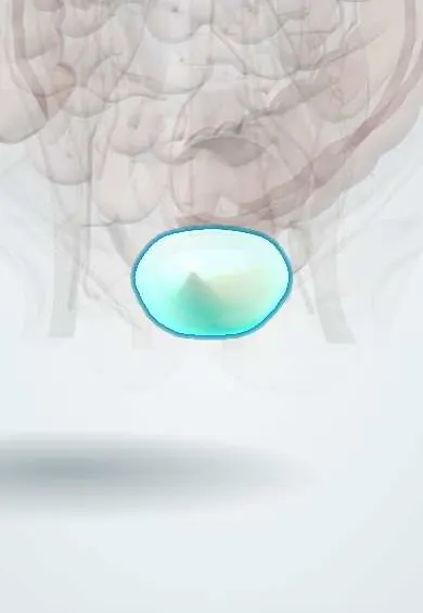
- Urinary bladder
- Membranous urethra
- Prostate
- Pubic symphysis
- Hip bone
- Prostatic urethra
- Spongy urethra
- External urethral sphincter
- Perineal membrane
- Urethra (male)
What it is
Rupture or contusion of the bladder, or partial or complete disruption of the urethra, most often linked to pelvic fractures. Bladder rupture is intraperitoneal or extraperitoneal; urethral injuries are posterior (membranous and prostatic urethra, with pelvic fracture) or anterior (bulbar and penile urethra, typically after a straddle fall).
Who gets it
Bladder injury occurs in a minority of pelvic fractures, and pelvic fracture urethral injury happens almost only in men. In Colombia these injuries follow mainly motorcycle crashes, run-over pedestrians and falls, and straddle injuries occur in work and cycling accidents.
Causes
- Pelvic ring fracture from road traffic crashes or crush injuries (posterior urethra, extraperitoneal bladder)
- Blow to a full bladder, e.g. seatbelt or kick (intraperitoneal rupture at the dome)
- Straddle falls onto a bar or beam (bulbar urethra)
- Penetrating wounds to the lower abdomen, perineum or buttocks
- Iatrogenic: catheterisation, pelvic surgery, endoscopic procedures
Risk factors
- Pelvic fracture, especially symphysis diastasis or pubic rami fractures
- Full bladder at the time of injury, often with alcohol intake
- Male sex (longer, more fixed urethra)
- Penile fracture during intercourse (anterior urethra)
Pathophysiology
A blow to a distended bladder raises its internal pressure and bursts the weakest part, the dome, leaking urine into the peritoneum. Pelvic fracture fragments and ligament shearing tear the extraperitoneal bladder wall or the membranous urethra where it crosses the urogenital diaphragm. Urine extravasation causes chemical peritonitis, infection and, through peritoneal reabsorption, rising creatinine.
Classification
- Bladder: contusion, extraperitoneal rupture (most common, with pelvic fracture) and intraperitoneal rupture (dome)
- Urethra: posterior (membranous or prostatic) versus anterior (bulbar or penile)
- Partial versus complete urethral disruption
Symptoms and signs
- Blood at the urethral meatus
- Visible haematuria (the classic sign of bladder rupture)
- Inability to void despite a full bladder
- Suprapubic pain and tenderness
- Perineal or scrotal bruising (butterfly pattern) or swelling
- High-riding or non-palpable prostate on rectal examination
Diagnosis
- Clinical
- Inspect the meatus and perineum in every pelvic trauma. A rectal examination can show a high-riding prostate, though it is unreliable. Urethral injury must be excluded before a urethral catheter is placed.
- Tests
- Urinalysis, haemoglobin, creatinine and electrolytes. Urinary drainage volume and colour are tracked.
- Imaging
- Retrograde urethrogram for suspected urethral injury. Retrograde cystography (plain or CT) with adequate bladder filling for visible haematuria with pelvic fracture; extravasation around the bowel loops means intraperitoneal rupture, a flame-shaped pattern in the pelvis means extraperitoneal rupture.
Differential diagnosis
- Renal or ureteric injury as the source of haematuria
- Pelvic haematoma compressing the bladder
- Acute urinary retention without injury
- Rectal injury
Goal of treatment
Drain urine safely, repair injuries that need it, and prevent stricture, incontinence and erectile dysfunction.
Treatment
- Surgical
- Intraperitoneal bladder rupture: surgical repair of the bladder wall with urinary catheter drainage.
- Conservative
- Uncomplicated extraperitoneal bladder rupture: urethral catheter drainage with follow-up cystography before removal.
- Surgical
- Complex extraperitoneal rupture (bladder neck, bone fragments, rectal or vaginal injury, or open pelvic fixation): surgical repair.
- Procedure
- Posterior urethral injury: early urinary drainage with a suprapubic catheter, sometimes primary endoscopic realignment in stable patients; definitive urethroplasty later.
- Surgical
- Anterior urethral injury: prompt repair of uncomplicated penetrating injuries; urinary drainage for straddle injuries with delayed reconstruction if a stricture forms.
- Follow-up
- Nursing priorities: do not attempt urethral catheterisation with blood at the meatus, record urine output and colour, care for the suprapubic tube, and watch for fever and abdominal signs.
Complications
- Urethral stricture
- Erectile dysfunction
- Urinary incontinence
- Urinary tract infection, pelvic abscess and urinary fistula
- Urinary ascites and peritonitis
Prognosis
Bladder injuries heal well when diagnosed and drained or repaired early. Pelvic fracture urethral injuries often lead to stricture needing reconstruction, and erectile dysfunction is common, so follow-up for at least a year is recommended.
Prevention
- Road safety measures
- Safe work practices at height
- No blind urethral catheterisation in pelvic trauma
Test yourself
Think of the answer, then tap the question to see it.
What must be done before a urethral catheter is placed in a pelvic trauma patient with blood at the meatus?
A retrograde urethrogram to rule out urethral injury.
Which type of bladder rupture always needs surgical repair?
Intraperitoneal rupture, usually at the dome after a blow to a full bladder.
How is an uncomplicated extraperitoneal bladder rupture usually managed?
With urethral catheter drainage and follow-up cystography.
Which urethral segment is injured in a straddle fall?
The bulbar (anterior) urethra, crushed against the pubic bone.
Related conditions
- Pelvic fracture
- Major trauma (polytrauma): ATLS primary survey
- Renal trauma
- Acute urinary retention
- Erectile dysfunction
- Urinary tract infection (acute cystitis)
- Peritonitis
- Acute kidney injury
Educational information, not medical advice. For any symptom, see a health professional.
Sources: Urotrauma Guideline - American Urological Association (American Urological Association, 2020); Kidney and uro-trauma: WSES-AAST guidelines (World Journal of Emergency Surgery, 2019).
Spotted a mistake?We are always improving for the community. Tell us what is wrong and our team will review it.Report a mistake
Representative model, for educational purposes only.