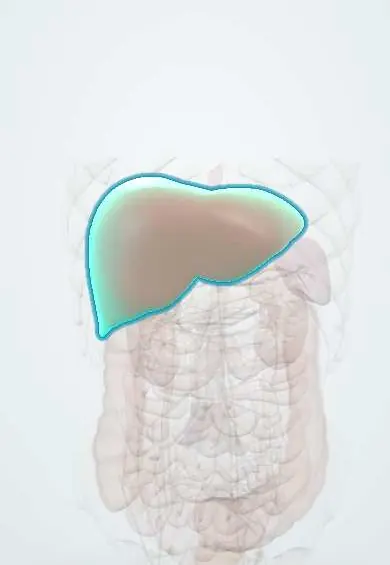
Conditions · Surgery and trauma
Liver injury !
Damage to the liver from a blow or a wound. It can bleed heavily, but most stable patients heal without surgery under close watch.
Updated 2026-11-01
Structures involved
Tap one to see what it is and open it in 3D.
- Liver
- Diaphragm
- Hepatic veins
- Inferior vena cava
- Portal vein
- Proper hepatic artery
- Common hepatic duct
- Gallbladder
- Spleen
- Kidneys
Why it happens
- Hemorrhage: petechiae, purpura, ecchymosis, hematoma, and what blood loss does
- Splanchnic and portal circulation
- Liver: lobule, acinus, hepatocytes, sinusoids, Kupffer cells
What it is
Laceration, haematoma or vascular disruption of the liver caused by blunt or penetrating trauma. The liver is the most frequently injured solid organ overall in abdominal trauma, especially in penetrating wounds, because of its size and position under the right lower ribs.
Who gets it
Liver injury appears in a large share of abdominal trauma cases. In Colombia, stab and gunshot wounds as well as motorcycle and car crashes produce many liver injuries in urban trauma centres; penetrating mechanisms are relatively more common than in high-income countries.
Causes
- Blunt trauma: road traffic crashes, falls, blows to the right upper quadrant
- Penetrating trauma: stab and gunshot wounds of the right upper quadrant or lower right chest
- Deceleration injury tearing the liver at its ligament attachments or hepatic veins
- Iatrogenic: liver biopsy or cardiopulmonary resuscitation
Risk factors
- Fractures of the lower right ribs
- Thoraco-abdominal stab wounds below the nipple line
- No seatbelt or motorcycle use
- Anticoagulant use or coagulopathy
- Pre-existing liver disease (large, fragile liver)
Pathophysiology
The liver is a large, vascular, relatively fixed organ, so crushing or shearing forces tear its parenchyma and vessels. Bleeding comes mainly from the portal and hepatic venous systems and from hepatic artery branches; injuries near the hepatic veins and retrohepatic vena cava are the most lethal. Bile leaks from torn ducts can cause biloma or bile peritonitis.
Classification
- AAST grades I–VI: from small subcapsular haematoma or capsular tear (I) to hepatic avulsion (VI)
- WSES classes combine the anatomic grade with haemodynamic status
- WSES I minor and II moderate: stable, AAST I–III
- WSES III severe: stable, AAST IV–V
- WSES IV: haemodynamically unstable, any grade
Symptoms and signs
- Right upper quadrant abdominal pain and tenderness
- Pain referred to the right shoulder (diaphragmatic irritation)
- Abdominal distension and guarding
- Signs of blood loss: tachycardia, pallor, low blood pressure
- Bruising or wounds over the right lower chest or upper abdomen
Diagnosis
- Clinical
- Suspect it with right upper quadrant or lower right chest trauma. The decisive question is haemodynamic status: unstable patients go straight to surgery; stable ones are staged with CT.
- Tests
- Serial haemoglobin, blood group and crossmatch, lactate and base deficit, coagulation tests, and liver enzymes. Repeated clinical examination is part of the work-up.
- Imaging
- FAST (focused assessment with sonography for trauma) at the bedside to look for free fluid. Contrast-enhanced CT in stable patients to grade the injury and detect active contrast extravasation (blush), pseudoaneurysm and associated injuries. Angiography for active arterial bleeding.
Differential diagnosis
- Splenic injury
- Right kidney injury
- Hollow organ (bowel or duodenal) injury
- Diaphragmatic rupture
- Rib fractures with chest wall pain only
Goal of treatment
Stop the bleeding, keep the patient alive and perfused, preserve liver tissue, and avoid unnecessary laparotomy in stable patients.
Treatment
- Acute
- ATLS resuscitation with early balanced blood products, massive transfusion protocol when needed, permissive hypotension until bleeding is controlled, and tranexamic acid early in bleeding patients.
- Conservative
- Non-operative management for haemodynamically stable patients without peritonitis or other injuries needing surgery, whatever the grade, in a centre with immediate access to surgery, ICU and blood.
- Procedure
- Angioembolisation for active arterial blush or pseudoaneurysm in stable or transiently responding patients.
- Surgical
- Damage control laparotomy for unstable patients: perihepatic packing, Pringle manoeuvre to slow inflow, control of bleeding vessels, and planned re-look once physiology is restored.
- Follow-up
- Nursing priorities during non-operative management: frequent vital signs, serial abdominal examination and haemoglobin, urine output, bed rest at first, and prompt escalation if instability or peritonitis develops.
Complications
- Delayed bleeding (the most common complication of non-operative management)
- Hepatic artery pseudoaneurysm and haemobilia
- Biloma, bile leak and bile peritonitis
- Liver abscess and liver necrosis
- Abdominal compartment syndrome
- Haemorrhagic shock and death
Prognosis
Most stable patients, including many with high-grade injuries, recover with non-operative management. Death is mainly linked to haemodynamic instability on arrival, injuries of the hepatic veins or retrohepatic vena cava, and associated injuries.
Prevention
- Seatbelt use
- Motorcycle safety
- Violence prevention
Test yourself
Think of the answer, then tap the question to see it.
What decides between operative and non-operative management of a liver injury?
Haemodynamic status. Stable patients without peritonitis or other operative injuries are managed non-operatively, whatever the grade.
What CT finding in a stable patient suggests angioembolisation?
Active arterial contrast extravasation (blush) or a pseudoaneurysm.
Which manoeuvre clamps the hepatoduodenal ligament to slow liver bleeding?
The Pringle manoeuvre, which occludes inflow through the hepatic artery and portal vein.
What do upper GI bleeding, jaundice and right upper quadrant pain after liver trauma suggest?
Haemobilia, usually from a ruptured hepatic artery pseudoaneurysm; it is treated with angioembolisation.
Related conditions
- Blunt abdominal trauma
- Penetrating trauma (stab and gunshot wounds)
- Splenic injury
- Major trauma (polytrauma): ATLS primary survey
- Hypovolaemic shock
- Rib fractures
- Peritonitis
- Renal trauma
Educational information, not medical advice. For any symptom, see a health professional.
Sources: Liver trauma: WSES 2020 guidelines (World Journal of Emergency Surgery, 2020).
Spotted a mistake?We are always improving for the community. Tell us what is wrong and our team will review it.Report a mistake
Representative model, for educational purposes only.