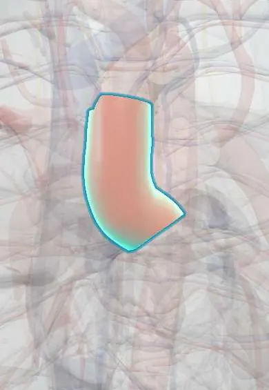
Transposition of the great arteries !
A birth heart defect where the aorta and pulmonary artery are swapped, so blue blood goes to the body. Needs urgent care and surgery.
Updated 2026-11-01
Structures involved
Tap one to see what it is and open it in 3D.
- Ascending aorta
- Pulmonary trunk
- Right ventricle
- Left ventricle
- Interatrial septum
- Interventricular septum
- Ligamentum arteriosum
- Aortic arch
- Pulmonary arteries
- Right atrium
- Left atrium
In embryology
- Outflow tract: the spiral aorticopulmonary septum and neural crest
- Transposition of the great arteries
- Maternal diabetes and obesity as teratogens
Why it happens
What it is
In dextro-transposition of the great arteries (d-TGA) the aorta arises from the right ventricle and the pulmonary artery from the left ventricle (ventriculoarterial discordance). The systemic and pulmonary circulations run in parallel instead of in series, so survival after birth depends on mixing of blood through a patent foramen ovale or atrial septal defect, a ventricular septal defect or the ductus arteriosus. It is the most common cyanotic congenital heart defect presenting in the first days of life.
Who gets it
About 5% of congenital heart disease, roughly 2–3 per 10,000 live births, with male predominance. In Colombia and Latin America, access to prenatal diagnosis and neonatal cardiac surgery varies by region, and delayed diagnosis worsens outcomes.
Causes
- Abnormal rotation and septation of the outflow tract (conotruncus) during embryonic development
- Usually not associated with a syndrome; rarely seen with chromosomal abnormalities
- Maternal pregestational diabetes is a recognised association
Risk factors
- Maternal diabetes
- Male sex
- Family history of congenital heart disease
- Maternal exposure to certain drugs (for example retinoids) or infections in pregnancy
Pathophysiology
Deoxygenated systemic venous blood returns to the right heart and is pumped back to the body through the aorta, while oxygenated pulmonary venous blood returns to the left heart and is pumped back to the lungs. Without a connection between the two circuits this is incompatible with life. When the ductus arteriosus closes in the first days, mixing falls and severe hypoxaemia and acidosis follow. Infants with an intact ventricular septum are the most cyanotic; those with a large VSD have better mixing but develop heart failure.
Classification
- d-TGA with intact ventricular septum (most common)
- d-TGA with ventricular septal defect
- d-TGA with VSD and left ventricular outflow (pulmonary) obstruction
- Congenitally corrected TGA (l-TGA): atrioventricular and ventriculoarterial discordance, not usually cyanotic
Symptoms and signs
- Central cyanosis in the first hours or days of life, not improved by oxygen
- Tachypnoea without marked respiratory distress
- Often no murmur when the septum is intact; single loud S2
- Heart failure signs (feeding difficulty, sweating, poor weight gain) in infants with a large VSD
- Rapid deterioration with acidosis and shock when the ductus closes
Diagnosis
- Clinical
- A cyanotic, otherwise vigorous newborn whose saturation does not improve with 100% oxygen (failed hyperoxia test) suggests cyanotic heart disease. Prenatal diagnosis on fetal echocardiography allows planned delivery at a cardiac centre.
- Tests
- Pulse oximetry screening of all newborns; arterial blood gas (low PaO2 that does not rise with oxygen, metabolic acidosis when ductus closes). ECG is often normal for age or shows right ventricular dominance.
- Imaging
- Echocardiography is diagnostic: parallel great arteries leaving the heart, aorta from the right ventricle and pulmonary artery from the left ventricle, size of the atrial communication, ductus and VSD, and coronary anatomy. Chest X-ray shows the classic «egg on a string» heart with a narrow mediastinum and increased pulmonary vascular markings.
Differential diagnosis
- Tetralogy of Fallot and other cyanotic heart defects (tricuspid atresia, truncus arteriosus, total anomalous pulmonary venous return)
- Persistent pulmonary hypertension of the newborn
- Respiratory disease of the newborn (respiratory distress syndrome, pneumonia)
- Methaemoglobinaemia
- Neonatal sepsis
Goal of treatment
Keep the ductus open and ensure adequate mixing of blood after birth, then perform definitive anatomical repair in the first weeks of life.
Treatment
- Acute
- Prostaglandin E1 (alprostadil) infusion to keep the ductus arteriosus open; watch for apnoea, fever and hypotension. Correct acidosis and support ventilation as needed.
- Procedure
- Balloon atrial septostomy (Rashkind procedure) to enlarge the atrial communication when mixing is inadequate.
- Surgical
- Arterial switch operation (Jatene), usually in the first two weeks of life: the great arteries are transected and reattached to the correct ventricles and the coronary arteries are reimplanted into the new aorta.
- Surgical
- Rastelli or related procedures for TGA with VSD and pulmonary outflow obstruction.
- Follow-up
- Nursing: continuous pulse oximetry and cardiac monitoring, temperature and glucose control, apnoea monitoring on prostaglandin, feeding and weight; lifelong follow-up by congenital cardiology.
Complications
- Severe hypoxaemia, acidosis, shock and death if untreated
- After arterial switch: coronary stenosis and ischaemia, supravalvular pulmonary or aortic stenosis, neo-aortic root dilation and regurgitation
- After older atrial switch operations (Mustard, Senning): systemic right ventricular failure, atrial arrhythmias, baffle obstruction
- Neurodevelopmental delay
Prognosis
Without treatment most infants die in the first months of life. After a timely arterial switch, long-term survival is excellent, though lifelong follow-up is needed for coronary, valvular and aortic problems.
Prevention
- Good preconception and pregnancy glucose control in women with diabetes
- Prenatal ultrasound with fetal echocardiography to plan delivery at a cardiac centre
- Universal newborn pulse oximetry screening for early detection
Test yourself
Think of the answer, then tap the question to see it.
Which ventricle gives rise to the aorta in d-TGA?
The right ventricle.
What is the classic chest X-ray appearance?
An «egg on a string» heart with a narrow mediastinum.
Which drug keeps the ductus arteriosus open before surgery?
Prostaglandin E1 (alprostadil).
Which maternal condition is associated with TGA?
Pregestational diabetes mellitus.
What is the definitive surgical repair?
The arterial switch (Jatene) operation with coronary reimplantation.
Related conditions
- Tetralogy of Fallot
- Truncus arteriosus, TAPVR and tricuspid atresia
- Ventricular septal defect
- Atrial septal defect
- Patent ductus arteriosus
- Persistent pulmonary hypertension of the newborn
- Heart failure
Educational information, not medical advice. For any symptom, see a health professional.
Sources: Transposition of the great arteries (MedlinePlus, US National Library of Medicine, 2026).
Spotted a mistake?We are always improving for the community. Tell us what is wrong and our team will review it.Report a mistake
Representative model, for educational purposes only.