Ventricular septal defect !
A ventricular septal defect is a hole, present from birth, in the wall between the heart's lower chambers; it is the commonest heart defect at birth.
Updated 2026-10-29
Structures involved
Tap one to see what it is and open it in 3D.
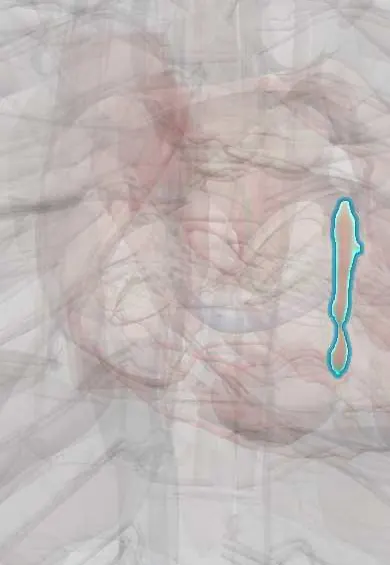
- Interventricular septum
- Left ventricle
- Right ventricle
- Pulmonary trunk
- Pulmonary arteries
- Left atrium
- Aortic right coronary cusp
- Tricuspid septal leaflet
- Bundle of His
In embryology
- 22q11.2 deletion (DiGeorge): the third and fourth pouches
- Ventricular septation and the endocardial cushions
- Ventricular septal defect
- Atrioventricular septal defect (endocardial cushion defect)
In the clinical exam
- Murmurs: timing, grade, location and radiation
- Valve murmurs: the table to know
- Dynamic auscultation: Valsalva, squatting, standing and handgrip
In physiology
Why it happens
What it is
An opening in the interventricular septum that lets blood flow between the ventricles, usually from left to right. It is the commonest congenital heart defect at birth, and many small defects close on their own.
Who gets it
The most frequent congenital heart defect in children, either alone or as part of complex defects such as tetralogy of Fallot. Many small muscular defects close in early childhood, so it is less common in adults.
Causes
- Incomplete septation of the ventricles during fetal development
- Chromosomal anomalies, such as trisomies 21, 18 and 13
- Part of complex defects, such as tetralogy of Fallot
- Rarely acquired: septal rupture after myocardial infarction or chest trauma
Risk factors
- Family history of congenital heart disease
- Maternal diabetes, rubella or alcohol use in pregnancy
- Exposure to some teratogenic drugs in pregnancy
- Prematurity
- Chromosomal syndromes
Pathophysiology
Left ventricular pressure is much higher than right, so blood crosses in systole into the right ventricle and pulmonary artery and returns to overload the left atrium and ventricle. Large defects cause heart failure in the first weeks, as pulmonary vascular resistance falls after birth. Untreated, pulmonary vascular disease can reverse the shunt (Eisenmenger syndrome).
Classification
- Perimembranous: the commonest type, near the tricuspid and aortic valves
- Muscular (trabecular): often multiple, many close spontaneously
- Outlet (doubly committed, supracristal): can let an aortic cusp prolapse, causing aortic regurgitation
- Inlet: part of the atrioventricular canal spectrum
- By size: restrictive (small) or non-restrictive (large)
Symptoms and signs
- Small defect: a loud, harsh holosystolic murmur at the lower left sternal border, often with a thrill, in a well child
- Large defect: fast breathing, sweating and tiredness during feeds
- Poor weight gain (failure to thrive)
- Recurrent chest infections
- Enlarged liver from heart failure
Diagnosis
- Clinical
- Harsh holosystolic murmur at the lower left sternal border; large defects add heart failure signs and a loud pulmonary component of S2.
- Tests
- ECG is normal in small defects and shows left or biventricular hypertrophy in large ones. Pulse oximetry checks for desaturation.
- Imaging
- Echocardiography with Doppler shows the site, size and gradient. Chest X-ray shows cardiomegaly and increased lung markings in large shunts. Catheterisation measures pulmonary vascular resistance in selected older patients.
Differential diagnosis
- Atrial septal defect
- Atrioventricular septal defect
- Patent ductus arteriosus
- Mitral regurgitation
- Tricuspid regurgitation
- Pulmonary valve stenosis
Goal of treatment
Control heart failure and support growth, close haemodynamically significant defects before irreversible pulmonary vascular disease, and prevent complications.
Treatment
- Follow-up
- Small restrictive defects are followed clinically and with echocardiography; many close spontaneously.
- Medical
- Infants with heart failure receive diuretics and sometimes ACE inhibitors, with calorie-dense feeding to support growth.
- Surgical
- Surgical patch closure on cardiopulmonary bypass for large defects with heart failure, poor growth, pulmonary hypertension or aortic cusp prolapse.
- Procedure
- Transcatheter device closure is an option for selected muscular and some perimembranous defects.
Complications
- Heart failure and failure to thrive
- Pulmonary hypertension and Eisenmenger syndrome
- Infective endocarditis
- Aortic regurgitation from cusp prolapse
- Heart block or arrhythmias after closure
Prognosis
Small defects have an excellent outlook and often close by themselves. After timely closure of large defects the long-term outlook is good; once Eisenmenger syndrome develops, the outlook is much worse.
Prevention
- Good dental hygiene to reduce endocarditis risk; antibiotic prophylaxis only in the high-risk situations named in current guidelines
- Rubella vaccination before pregnancy
Test yourself
Think of the answer, then tap the question to see it.
What murmur is typical of a ventricular septal defect?
A harsh holosystolic murmur at the lower left sternal border, often with a thrill.
What is the commonest anatomical type of ventricular septal defect?
The perimembranous defect.
Why does a large ventricular septal defect cause heart failure weeks after birth rather than at birth?
Pulmonary vascular resistance falls over the first weeks, so the left-to-right shunt grows.
Is a louder murmur a sign of a larger ventricular septal defect?
No. Small restrictive defects often have the loudest murmurs because the jet is fast.
Which valve problem can complicate an outlet ventricular septal defect?
Aortic regurgitation, from prolapse of an aortic cusp into the defect.
Related conditions
- Atrial septal defect
- Tetralogy of Fallot
- Patent ductus arteriosus
- Heart failure
- Myocardial Infarction (Heart Attack)
- Infective endocarditis
- Aortic regurgitation
- Mitral regurgitation
Educational information, not medical advice. For any symptom, see a health professional.
Sources: Ventricular Septal Defect (StatPearls (NCBI Bookshelf), 2024).
Spotted a mistake?We are always improving for the community. Tell us what is wrong and our team will review it.Report a mistake
Representative model, for educational purposes only.