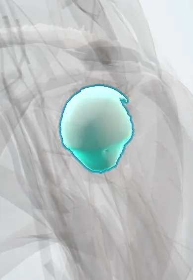
Humeral head cartilage !
The humeral head cartilage is the smooth layer covering the ball of the shoulder; it lets the ball glide in the socket with almost no friction.
Official name: Articular cartilage of the humeral head
Opens in the Durgical app, no sign-up needed.
What it is
The smooth layer covering the ball of the shoulder.
What it does
Lets the ball glide in the socket with almost no friction.
Key facts
- Where
- Humeral head (anatomical neck margin up)
- Function
- Hyaline cartilage; smooth, low-friction joint surface; wears in osteoarthritis
Relations
- Deep
- Subchondral bone of the humeral head; at its edge, the anatomical neck of the humerus, where the shoulder capsule attaches
- Medial
- Glenoid cartilage and the glenoid labrum, across the joint space
- Above
- Long biceps tendon arching over the head inside the joint, then the supraspinatus tendon above the capsule
- In front
- Subscapularis tendon, separated from it by the capsule and the middle glenohumeral ligament (MGHL)
- Behind
- Infraspinatus tendon and teres minor tendon, wrapping the back of the head
How to find it
It cannot be felt. At shoulder arthroscopy it is seen from the posterior portal, with the bare area and any Hill-Sachs dent on its back; in open surgery it comes into view through a deltopectoral approach once the subscapularis is detached or split.
On imaging
A true AP (Grashey) view shows the joint space as a clear gap and the outline of the head; an AP in internal rotation or a Stryker notch view shows a Hill-Sachs dent. MRI shows cartilage thinning, defects and bone oedema; CT measures the size and position of a Hill-Sachs lesion before surgery.
Common problems
- Shoulder osteoarthritis: the cartilage wears thin, causing pain, grinding and stiffness.
- Cuff tear arthropathy: a large old rotator cuff tear lets the ball ride up and wear out.
- Damage after repeated dislocations.
How it is diagnosed
X-rays show the narrowed joint space and bone spurs; CT plans surgery; MRI checks the rotator cuff.
Treatment
- Activity changes, pain medicines, physical therapy and sometimes injections.
- Total shoulder replacement when the cuff is healthy; reverse replacement when the cuff is torn.
Clinical notes
- A large Hill-Sachs dent can catch ('engage') on the front of the glenoid in abduction and external rotation; together with glenoid bone loss it guides the choice of surgery, such as remplissage (filling the dent with infraspinatus) or a bone block
- Avascular necrosis and collapse of the head can follow anatomical neck or four-part proximal humerus fractures, which cut the supply from the circumflex humeral arteries
- Anatomic total shoulder arthroplasty resurfaces the head and glenoid when the rotator cuff works; a reverse prosthesis is chosen when the cuff has failed
Development
The head ossifies from its own secondary centre in infancy and later fuses with the centres of the greater and lesser tubercles; the proximal humeral physis beneath it closes in late adolescence and can be mistaken for a fracture line on X-ray.
Related conditions
Test yourself
Think of the answer, then tap the question to see it.
What is a Hill-Sachs lesion of the humeral head cartilage?
A dent in the back, upper and outer humeral head made by the glenoid rim in an anterior dislocation.
Where is the humeral head cartilage thickest?
At the centre of the head, thinner at the edge: the reverse of the glenoid cartilage.
Why can avascular necrosis of the humeral head follow anatomical neck or four-part fractures?
These fractures cut the supply from the circumflex humeral arteries.
When is a reverse shoulder prosthesis chosen instead of an anatomic one?
When the rotator cuff has failed; anatomic replacement resurfaces head and glenoid when the cuff works.
Educational information, not medical advice. For any symptom, see a health professional.
Its names
- Official Latin (TA2)
- cartilago articularis capitis humeri TA2 382
- Official Spanish (DeCS)
- Cartílago articular de la cabeza del húmero
- English (TA2)
- Articular cartilage of the humeral head
Related
Sources: Terminologia Anatomica (TA2); Anatomy, Shoulder and Upper Limb, Glenohumeral Joint; Anatomy, Shoulder and Upper Limb, Shoulder.
Spotted a mistake?We are always improving for the community. Tell us what is wrong and our team will review it.Report a mistake
Representative model, for educational purposes only.