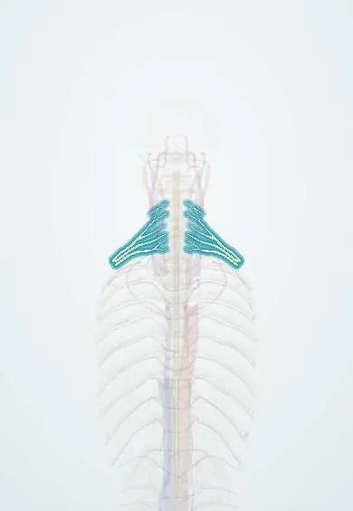
Brachial plexus injury !
A brachial plexus injury is damage to the network of nerves from the neck to the arm, usually by stretching; it causes weakness and numbness of the arm.
Updated 2026-10-29
Structures involved
Tap one to see what it is and open it in 3D.
- Brachial plexus roots
- Plexus trunks
- Medial cord
- Plexus divisions
- Lateral cord
- Posterior cord
- Musculocutaneous nerve
- Axillary nerve
- Suprascapular nerve
Why it happens
- Upper versus lower motor neuron lesions
- Peripheral nerve: endoneurium, perineurium, epineurium; myelin
- Nerve injury and regeneration (Wallerian degeneration)
What it is
Damage to the network of nerves formed by the C5–T1 roots that supplies the upper limb, usually from traction. The classic patterns are Erb palsy (upper plexus, C5–C6) and Klumpke palsy (lower plexus, C8–T1).
Who gets it
In adults most injuries follow high-energy trauma, above all motorcycle crashes, a major cause in Colombia and Latin America. In newborns obstetric injury follows difficult deliveries, especially shoulder dystocia.
Causes
- Traction from forced separation of the head and shoulder (motorcycle falls, falls onto the shoulder)
- Forced arm abduction above the head, stretching the lower plexus (grabbing a support during a fall)
- Obstetric traction during delivery (shoulder dystocia, breech delivery)
- Penetrating wounds, clavicle fracture, shoulder dislocation, tumours or radiation
Risk factors
- Motorcycle riding
- Contact sports (stingers or burners)
- Macrosomia, maternal diabetes and shoulder dystocia (obstetric injury)
- Previous radiation to the axilla or neck
Pathophysiology
Traction can stretch the nerve (neurapraxia), break the axons (axonotmesis), rupture the nerve (neurotmesis) or tear the roots from the spinal cord (avulsion), which cannot repair itself. Upper plexus injury (C5–C6) weakens shoulder abductors, external rotators and elbow flexors. Lower plexus injury (C8–T1) weakens the intrinsic hand muscles; T1 avulsion can damage the sympathetic chain and cause Horner syndrome.
Classification
- Erb-Duchenne (upper, C5–C6, sometimes C7): waiter's tip posture
- Klumpke (lower, C8–T1): claw hand, sometimes with Horner syndrome
- Total (pan-plexus) injury: flail, numb arm
- Preganglionic (root avulsion) versus postganglionic (rupture or stretch)
- Seddon or Sunderland nerve injury grades
Symptoms and signs
- Erb: arm hanging adducted and internally rotated, elbow extended, forearm pronated, wrist flexed (waiter's tip)
- Klumpke: claw hand with weak intrinsic hand muscles and numbness on the ulnar forearm and hand
- Ptosis, miosis and anhidrosis on the same side if T1 is involved (Horner)
- Burning (neuropathic) pain in the arm, typical of root avulsion
- In newborns: reduced movement of one arm and an asymmetric Moro reflex
Diagnosis
- Clinical
- Detailed motor and sensory mapping by root, trunk and cord. Weak serratus anterior (winged scapula), rhomboids or a Horner sign point to a preganglionic lesion. Tinel sign along the plexus is followed over time.
- Tests
- Electromyography and nerve conduction studies, usually after a few weeks; preserved sensory potentials in a numb area suggest root avulsion.
- Imaging
- X-rays of the cervical spine, shoulder, clavicle and chest (a raised hemidiaphragm suggests phrenic involvement). MRI or CT myelography looks for pseudomeningoceles from root avulsion; angiography if vascular injury is suspected.
Differential diagnosis
- Spinal cord injury
- Cervical radiculopathy
- Isolated peripheral nerve injury (axillary, radial, suprascapular)
- Rotator cuff tear or shoulder dislocation
- Neuralgic amyotrophy (Parsonage-Turner syndrome)
- Clavicle or humerus fracture causing pseudoparalysis (in newborns)
Goal of treatment
Recover the most useful functions (elbow flexion, shoulder stability, hand sensation), control pain and prevent contractures.
Treatment
- Acute
- In trauma, initial care follows trauma protocols: airway, bleeding control, cervical spine protection and repair of vascular injuries.
- Follow-up
- Closed stretch injuries are usually observed with serial examination and electromyography, because many recover over months.
- Surgical
- Surgery for open injuries, avulsions or failed recovery: nerve repair, nerve grafts, nerve transfers and later tendon or free muscle transfers; timing matters because muscles lose reinnervation potential.
- Rehabilitation
- Physiotherapy and occupational therapy to keep joints mobile, prevent contractures and retrain function; in babies, parents learn gentle range-of-motion exercises.
- Medical
- Neuropathic pain medications such as anticonvulsants or antidepressants of the neuropathic pain classes.
Complications
- Permanent weakness or paralysis of the arm
- Chronic neuropathic pain
- Joint contractures and shoulder deformity (internal rotation contracture in children)
- Limb length or size difference in obstetric palsy
- Psychological and occupational impact
Prognosis
Depends on the level and type of injury. Most neonatal Erb palsies recover well in the first months. Upper plexus injuries recover better than lower ones, and root avulsions do not recover without surgery.
Prevention
- Helmets, protective gear and road safety for motorcyclists
- Recognition and management of shoulder dystocia in delivery rooms
Test yourself
Think of the answer, then tap the question to see it.
Which roots are injured in Erb palsy, and what posture does it produce?
C5–C6 (upper trunk). The arm hangs adducted and internally rotated, with the elbow extended and the forearm pronated: the waiter's tip posture.
Which roots are injured in Klumpke palsy, and what are its main signs?
C8–T1 (lower trunk). Claw hand with weak intrinsic muscles, ulnar-side numbness and, if T1 is involved, Horner syndrome.
Why does Horner syndrome suggest a severe brachial plexus injury?
It means the T1 root is injured near the spinal cord, damaging sympathetic fibres; this often indicates a preganglionic root avulsion.
What mechanism causes most adult brachial plexus injuries in Latin America?
High-energy traction injury, typically a motorcycle crash that forces the head and shoulder apart.
Related conditions
- Spinal cord injury
- Clavicle fracture
- Shoulder dislocation
- Thoracic outlet syndrome
- Cervical radiculopathy
- Proximal humerus fracture
- Winged scapula
- Axillary nerve injury
- Rotator cuff tear
Educational information, not medical advice. For any symptom, see a health professional.
Sources: Brachial Plexus Injuries (StatPearls (NCBI Bookshelf), 2026).
Spotted a mistake?We are always improving for the community. Tell us what is wrong and our team will review it.Report a mistake
Representative model, for educational purposes only.