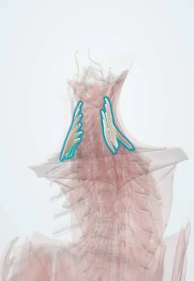
Scalenes !
The scalenes are three muscles on the side of the neck, from the neck vertebrae to the first two ribs; they bend the neck and lift the ribs.
Official name: Scalenus anterior muscle
Opens in the Durgical app, no sign-up needed.
What it is
Three muscles on the side of the neck, from the neck vertebrae to the first two ribs.
What it does
Bend the neck to the side and lift the upper ribs when you breathe hard. The brachial plexus and subclavian artery pass between the front and middle scalenes (thoracic outlet).
Key facts
- Origin
- Cervical transverse processes: anterior C3–C6, middle C2–C7, posterior C4–C6
- Insertion
- Anterior: scalene tubercle of 1st rib; middle: 1st rib behind subclavian groove; posterior: 2nd rib
- Action
- Lateral flexion of the neck; raise 1st–2nd ribs in forced inspiration; anterior also flexes the neck
- Nerve
- Ventral rami of cervical nerves: anterior C4–C6, middle C3–C8, posterior C6–C8 (varies)
- Blood supply
- Ascending cervical artery (from inferior thyroid); transverse cervical artery
Relations
- In front
- Anterior scalene: phrenic nerve on its surface, subclavian vein, transverse cervical and suprascapular arteries; on the left the thoracic duct arches in front; sternocleidomastoid over it
- Contents
- Interscalene triangle: roots and trunks of the brachial plexus and the subclavian artery
- Behind
- Levator scapulae behind the middle and posterior scalenes; the dorsal scapular nerve and the roots of the long thoracic nerve pierce the middle scalene
- Medial
- Longus colli, with the vertebral artery rising between it and anterior scalene to the C6 transverse foramen
- Below
- First rib (scalene tubercle, with the groove for the subclavian artery just behind) and second rib; the dome of the pleura just below and medial
How to find it
Just above the clavicle, behind the clavicular head of sternocleidomastoid, anterior scalene is felt as a firm band that tightens on a deep breath in. The interscalene groove between anterior and middle scalenes, at the level of the cricoid cartilage (C6), is where the brachial plexus is found for a block.
Examination
- Adson test: neck extended and head turned toward the tested side (some texts: away), with a deep breath in while the radial pulse is felt; a fading pulse with symptoms suggests subclavian artery compression at the interscalene triangle, though false positives are common
On imaging
Ultrasound shows anterior and middle scalene with the brachial plexus roots between them as round dark dots (the 'traffic light' view) for an interscalene block, and the phrenic nerve on anterior scalene. In thoracic outlet syndrome, neck radiographs look for a cervical rib or a long C7 transverse process; CT or MR angiography with the arm raised can show compression of the subclavian vessels.
Clinical notes
- Thoracic outlet syndrome: the brachial plexus (most cases are neurogenic) or the subclavian vessels are compressed in the interscalene triangle, the costoclavicular space or under pectoralis minor; a cervical rib or fibrous band is a classic cause
- Surgical decompression of the outlet removes part of the first rib and, often, divides anterior scalene
- Anterior scalene is the key landmark at the root of the neck: subclavian vein in front, subclavian artery behind, phrenic nerve on its surface
Anatomical variants
- Anterior scalene attachments vary, and it occasionally lies behind the subclavian artery instead of in front; parts of the brachial plexus may pierce it; a small scalenus minimus is present in some people
Development
They develop from the ventral part of the cervical myotomes, hence their ventral ramus supply. The costal element of C7 normally stays small, but it can grow into a cervical rib or a fibrous band that narrows the interscalene triangle and the space above the first rib.
Related conditions
Test yourself
Think of the answer, then tap the question to see it.
What passes through the interscalene triangle?
The roots and trunks of the brachial plexus and the subclavian artery, between the anterior and middle scalenes and the first rib.
Which structures lie in front of and on the anterior scalene?
The subclavian vein passes in front; the phrenic nerve runs down on its surface, from lateral to medial.
Why does an interscalene brachial plexus block cause hemidiaphragm paralysis?
It nearly always also blocks the phrenic nerve, which lies on the anterior scalene.
Where do the scalenes insert?
Anterior: scalene tubercle of the 1st rib; middle: 1st rib behind the subclavian groove; posterior: 2nd rib.
What is the classic bony cause of thoracic outlet syndrome at the scalenes?
A cervical rib or fibrous band narrowing the interscalene triangle; most cases are neurogenic.
Educational information, not medical advice. For any symptom, see a health professional.
Its names
- Official Latin (TA2)
- musculus scalenus anterior; musculus scalenus medius; musculus scalenus posterior TA2 2152
- Official Spanish (DeCS)
- Músculos escalenos DeCS D009334
- English (TA2)
- Scalenus anterior muscle
- Also called
- anterior scalene muscle · middle scalene muscle · posterior scalene muscle · musculus scalenus ventralis
Related
- Brachial plexus roots
- Neck nerve roots
- Axis (C2)
- C3 vertebra
- C4 vertebra
- C5 vertebra
- C6 vertebra
- C7 vertebra
Sources: Terminologia Anatomica (TA2); DeCS; Anatomy, Head and Neck, Scalenus Muscle; Anatomy of the Human Body (20th ed.): Development of the Muscles (Lea & Febiger (Gray's Anatomy, 1918), 1918) · adapted, Public domain; Anatomy of the Human Body (20th ed.): The Cervical Vertebrae (Lea & Febiger (Gray's Anatomy, 1918), 1918) · adapted, Public domain; Anatomy of the Human Body (20th ed.): The Costal Cartilages (Lea & Febiger (Gray's Anatomy, 1918), 1918) · adapted, Public domain.
Spotted a mistake?We are always improving for the community. Tell us what is wrong and our team will review it.Report a mistake
Representative model, for educational purposes only.