Costochondritis !
Costochondritis is inflammation of the cartilage where the ribs join the breastbone, causing chest pain that hurts more when the spot is pressed.
Updated 2026-10-29
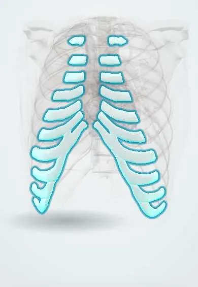
Structures involved
Tap one to see what it is and open it in 3D.
What it is
Inflammation of the cartilage where the ribs join the sternum, causing sharp anterior chest pain that is reproduced by pressing on the joint. It is a diagnosis of exclusion once cardiac and other serious causes are ruled out.
Who gets it
A frequent cause of chest pain in primary care and emergency departments, especially in women, adolescents and young adults.
Causes
- Usually idiopathic
- Repetitive minor trauma or unusual exertion
- Severe or persistent coughing
- After respiratory infections
- Rarely infection after sternotomy or injection drug use
Risk factors
- Female sex
- Age under 40
- Strenuous upper body exercise or heavy lifting
- Recent respiratory infection with cough
- Fibromyalgia
Pathophysiology
Low-grade inflammation of the costochondral and costosternal joints, usually ribs 2 to 5, sensitises the joint, so movement, deep breathing and pressure provoke pain. Unlike Tietze syndrome, there is no visible swelling.
Symptoms and signs
- Sharp or aching anterior chest pain, often on the left
- Pain reproduced by pressing on the costochondral junctions
- Pain worse with deep breathing, coughing or moving the arms and trunk
- Usually several adjacent junctions, without swelling
Diagnosis
- Clinical
- Reproducible point tenderness at the costochondral junctions with a normal examination otherwise. It is diagnosed only after cardiac and other serious causes are reasonably excluded; tenderness alone does not rule out heart disease.
- Tests
- ECG and troponin when the history or risk factors suggest a cardiac cause; inflammatory markers if infection or systemic disease is suspected.
- Imaging
- Chest X-ray when lung or bone disease is possible; imaging is normal in costochondritis.
Differential diagnosis
- Acute coronary syndrome or stable angina
- Pericarditis
- Pulmonary embolism
- Pneumothorax
- Gastro-oesophageal reflux
- Tietze syndrome or rib fracture
Goal of treatment
Relieve pain, reassure once serious causes are excluded and return the patient to normal activity.
Treatment
- Conservative
- Explanation and reassurance, relative rest and avoiding activities that provoke the pain; local heat.
- Medical
- Simple analgesics and non-steroidal anti-inflammatory drugs, oral or topical, if there are no contraindications.
- Rehabilitation
- Gentle chest wall stretching and physiotherapy for persistent cases.
- Follow-up
- Review if pain changes or warning signs appear; local injection is rarely used for refractory cases.
Complications
- Missed cardiac or pulmonary diagnosis if evaluation is incomplete
- Chronic or recurrent chest pain
- Anxiety about heart disease
Prognosis
Benign and self-limiting; most cases resolve in weeks to months, though some recur.
Test yourself
Think of the answer, then tap the question to see it.
What finding supports costochondritis on examination?
Pain reproduced by pressing on the costochondral or costosternal junctions, usually ribs 2 to 5.
Why is costochondritis called a diagnosis of exclusion?
Because cardiac, pulmonary and other serious causes of chest pain must be excluded first; chest wall tenderness does not rule them out.
How is costochondritis different from Tietze syndrome?
Tietze syndrome has visible swelling of the joint, usually ribs 2 to 3; costochondritis has no swelling.
What drug class is usually used for costochondritis pain?
Non-steroidal anti-inflammatory drugs, oral or topical, plus simple analgesics.
Related conditions
- Stable angina
- Myocardial Infarction (Heart Attack)
- Gastro-oesophageal reflux disease (GERD)
- Pulmonary embolism
- Pneumothorax
- Acute pericarditis
- Rib fractures
Educational information, not medical advice. For any symptom, see a health professional.
Sources: Costochondritis (StatPearls (NCBI Bookshelf), 2024).
Spotted a mistake?We are always improving for the community. Tell us what is wrong and our team will review it.Report a mistake
Representative model, for educational purposes only.