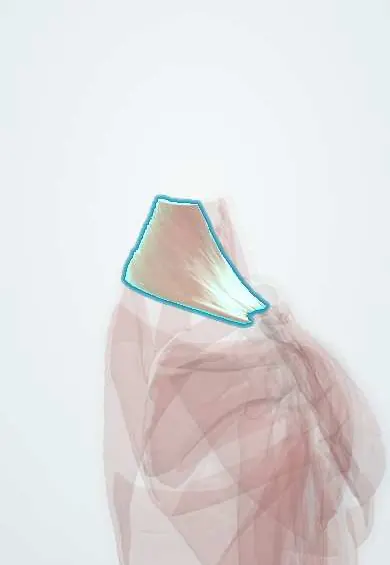
Upper trapezius !
The upper trapezius runs from the neck to the top of the shoulder; it shrugs the shoulder and helps rotate the shoulder blade upward.
Official name: Trapezius, upper (descending) part
Opens in the Durgical app, no sign-up needed.
What it is
The upper part of the large diamond-shaped muscle of the upper back and neck.
What it does
Shrugs the shoulder and helps rotate the shoulder blade upward.
Key facts
- Origin
- External occipital protuberance, superior nuchal line, nuchal ligament
- Insertion
- Lateral third of the clavicle
- Action
- Elevates and upwardly rotates the scapula; extends and side-bends the neck
- Nerve
- Accessory nerve (CN XI); C3–C4 (proprioception)
- Blood supply
- Transverse cervical artery, occipital artery
Relations
- Superficial
- Skin of the nape and top of the shoulder; the investing layer of deep cervical fascia splits to enclose it
- Deep
- Splenius capitis, semispinalis capitis and levator scapulae; the accessory nerve and the superficial branch of the transverse cervical artery run on its deep surface
- In front
- The posterior triangle of the neck, with the sternocleidomastoid as its front border; the triangle holds CN XI, the trunks of the brachial plexus and the lymph nodes of the posterior neck
- Below
- The lateral third of the clavicle, where it inserts opposite the origin of the front deltoid; below the C7 level it continues as the middle trapezius
How to find it
With the person seated and the examiner behind, a shrug against downward pressure on both shoulders makes it stand out as the sloping ridge from the base of the skull to the outer end of the clavicle. Comparing both sides shows a droop or loss of bulk.
Examination
- Resisted shrug: weakness or a lower, flatter shoulder on one side points to an accessory nerve (CN XI) lesion; normal head turning with a weak shrug places the lesion below the branch to the sternocleidomastoid, as in the posterior triangle
On imaging
Ultrasound and MRI show it as a thin sheet over the back of the neck and shoulder. After accessory nerve injury, often from surgery or a biopsy in the posterior triangle, CT or MRI shows the muscle thin and fatty compared with the other side. Ultrasound can follow the nerve in the posterior triangle; electromyography confirms denervation.
Clinical notes
- The accessory nerve can be cut during a lymph node biopsy or a neck dissection in the posterior triangle: the shoulder droops and aches, the arm cannot be raised fully to the side and the scapula wings laterally
- Neck dissections that spare CN XI were developed to avoid this painful 'shoulder syndrome'
- Scapular dyskinesis often shows an overactive upper trapezius (the shoulder hikes up as the arm rises) with weak lower trapezius and serratus anterior
Anatomical variants
- Rare: absence or underdevelopment of part of the muscle, and accessory slips
Development
The trapezius and sternocleidomastoid grow from one premuscle mass in the occipital region, just behind the last pharyngeal arch; the accessory nerve enters it early and is carried down as the mass spreads to the shoulder girdle and spine.
Related conditions
Test yourself
Think of the answer, then tap the question to see it.
What nerve supplies the upper trapezius, and how is it tested at the bedside?
The accessory nerve (CN XI); by a resisted shoulder shrug.
Why is the accessory nerve at risk in the posterior triangle of the neck?
It crosses the triangle just under the skin and fascia to reach the upper trapezius; a lymph node biopsy or neck dissection can cut it.
A patient has a weak shrug but normal head turning against resistance. Where is the accessory nerve lesion?
Below the branch to the sternocleidomastoid, as in the posterior triangle of the neck.
Educational information, not medical advice. For any symptom, see a health professional.
Its names
- Official Latin (TA2)
- pars descendens musculi trapezii TA2 2227
- Official Spanish (DeCS)
- Porción descendente del músculo trapecio
- English (TA2)
- Trapezius, upper (descending) part
Related
Sources: Terminologia Anatomica (TA2); Anatomy, Back, Trapezius; Anatomy of the Human Body (20th ed.): Development of the Muscles (Lea & Febiger (Gray's Anatomy, 1918), 1918) · adapted, Public domain.
Spotted a mistake?We are always improving for the community. Tell us what is wrong and our team will review it.Report a mistake
Representative model, for educational purposes only.