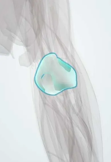
Elbow dislocation !
An elbow dislocation is when the forearm bones come out of the elbow joint, nearly always backward; it is the second most dislocated large joint in adults.
Updated 2026-10-29
Structures involved
Tap one to see what it is and open it in 3D.
- Elbow capsule
- Ulna
- Humerus
- UCL (elbow)
- Radius
- Radial head cartilage
- Brachial artery
- Median nerve
- Ulnar nerve
What it is
Loss of contact between the humerus and the ulna and radius at the elbow, nearly always with the forearm bones displaced backwards (posterior dislocation). It is the second most common major joint dislocation in adults after the shoulder.
Who gets it
Typical in young adults and adolescents after falls during sport, and in older people after simple falls. In children, supracondylar fractures are more common than dislocations after a similar fall.
Causes
- Fall onto an outstretched hand with the elbow extended
- Combined axial load, valgus and supination of the forearm
- High-energy trauma such as traffic accidents
Risk factors
- Contact sports, gymnastics, skating and cycling
- Falls in older adults
- Ligamentous laxity
- Previous elbow dislocation
Pathophysiology
Injury usually progresses from lateral to medial (Horii circle): the lateral ulnar collateral ligament fails first, then the capsule, and finally the medial collateral ligament. Simple dislocations involve only soft tissues; complex ones include fractures. The brachial artery and the median and ulnar nerves cross the elbow and can be stretched or trapped.
Classification
- By direction: posterior or posterolateral (most common), anterior, medial, lateral, divergent
- Simple (no fracture) versus complex (with fracture)
- Terrible triad: elbow dislocation with radial head fracture and coronoid process fracture, highly unstable
- Other complex patterns: dislocation with medial epicondyle fracture in adolescents
Symptoms and signs
- Severe pain and obvious deformity after a fall
- Elbow held in partial flexion, unable to move
- Prominent olecranon at the back of the elbow
- Swelling and later bruising
Diagnosis
- Clinical
- Deformity with loss of the normal triangle between the epicondyles and the olecranon. Clinicians check radial and ulnar pulses, capillary refill, and median, ulnar and radial nerve function before and after reduction, and examine the shoulder and wrist.
- Tests
- Not usually needed. Pulse oximetry or Doppler of the hand if circulation is in doubt.
- Imaging
- X-rays before and after reduction in two views to confirm direction, look for fractures (radial head, coronoid, epicondyles) and confirm a concentric reduction. CT for complex fractures; angiography if arterial injury is suspected.
Differential diagnosis
- Supracondylar humerus fracture (especially in children)
- Radial head or olecranon fracture
- Monteggia fracture-dislocation
- Radial head subluxation in young children
- Isolated radial head dislocation
Goal of treatment
Reduce the joint promptly, protect the artery and nerves, restore a stable elbow and recover motion while avoiding stiffness.
Treatment
- Acute
- Prompt closed reduction under analgesia or sedation, followed by checks of pulses and nerves and post-reduction X-rays.
- Conservative
- Simple stable dislocations: short immobilisation in a splint for comfort, then early protected movement to avoid stiffness.
- Surgical
- Surgery for complex dislocations (terrible triad: fixation or replacement of the radial head, coronoid fixation and lateral ligament repair), irreducible or unstable joints, and arterial injury.
- Rehabilitation
- Physiotherapy to regain flexion, extension and forearm rotation.
Complications
- Stiffness and loss of full extension (the most common)
- Recurrent instability
- Heterotopic ossification
- Brachial artery injury
- Median or ulnar nerve injury, sometimes with nerve trapped in the joint
- Compartment syndrome and post-traumatic arthritis
Prognosis
Simple dislocations usually do well with early movement, though a small loss of extension is common. Complex injuries, especially the terrible triad, carry more risk of stiffness, instability and arthritis.
Prevention
- Protective technique and equipment in sport
- Fall prevention in older adults
Test yourself
Think of the answer, then tap the question to see it.
What is the most common direction of elbow dislocation?
Posterior (or posterolateral): the ulna and radius move backwards relative to the humerus.
What is the terrible triad of the elbow?
Elbow dislocation with a radial head fracture and a coronoid process fracture; it is highly unstable and usually treated surgically.
Which artery and nerves must be checked before and after reducing an elbow dislocation?
The brachial artery (radial and ulnar pulses) and the median, ulnar and radial nerves.
Which ligament usually fails first in a posterolateral elbow dislocation?
The lateral ulnar collateral ligament, at the start of the lateral-to-medial injury sequence (Horii circle).
What is the most common complication after a simple elbow dislocation?
Stiffness, especially loss of full extension, which is why early movement is encouraged.
Related conditions
- Supracondylar humerus fracture
- Acute compartment syndrome
- Cubital tunnel syndrome (ulnar neuropathy at the elbow)
- Shoulder dislocation
- Distal radius fracture (Colles)
- Radial head subluxation (pulled elbow)
- Olecranon bursitis
- Humeral shaft fracture
Educational information, not medical advice. For any symptom, see a health professional.
Sources: Posterior Elbow Dislocation (StatPearls (NCBI Bookshelf), 2023).
Spotted a mistake?We are always improving for the community. Tell us what is wrong and our team will review it.Report a mistake
Representative model, for educational purposes only.