Conditions · Pediatrics and neonatology
Anorectal malformations (imperforate anus) !
Birth defects in which the anus is missing, blocked or misplaced, often with a fistula to the urinary tract or vagina. Surgery repairs them.
Updated 2026-11-01
Structures involved
Tap one to see what it is and open it in 3D.
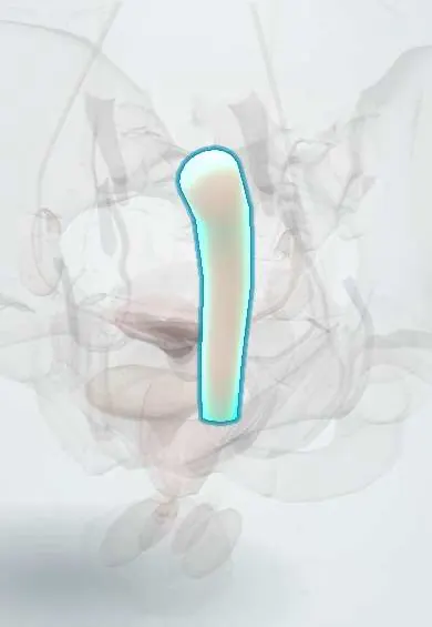
- Anal canal
- Rectum
- External anal sphincter
- Puborectalis
- Perineal body
- Sacrum
- Prostatic urethra
- Urinary bladder
Why it happens
What it is
A spectrum of congenital defects of the distal rectum and anus in which the anal opening is absent, narrowed or displaced, often with an abnormal connection (fistula) to the perineum, urethra, bladder neck, vestibule or vagina.
Who gets it
About 1 in 4,000 to 5,000 live births, slightly more often in boys. Around half of affected children have another anomaly, most often urinary, spinal, cardiac or as part of VACTERL association; it is also more frequent in Down syndrome. In Colombia it is managed by pediatric surgery in referral hospitals.
Causes
- Abnormal division of the cloaca by the urorectal septum in the first weeks of embryonic life
- Most cases sporadic; occasional familial or syndromic cases (VACTERL, Down syndrome, Currarino syndrome)
Risk factors
- Other congenital anomalies, especially VACTERL association
- Down syndrome
- Family history of anorectal malformation
Pathophysiology
If the urorectal septum fails to separate the cloaca properly, the rectum ends blindly or opens at an abnormal site. The level of the rectal pouch relative to the pelvic floor muscles (levator ani and puborectalis) and the external sphincter determines the type of fistula and the chances of later bowel control. Abnormal sacral development and tethered spinal cord, which often coexist, further impair continence.
Classification
- Boys: perineal fistula (low); rectourethral bulbar or prostatic fistula; rectobladder neck fistula (highest)
- Girls: perineal fistula; vestibular fistula (most common in girls); cloaca (rectum, vagina and urethra share one channel)
- Imperforate anus without fistula (common in Down syndrome) and rectal atresia or stenosis
- Older scheme: low versus high lesions
Symptoms and signs
- No anal opening, or an opening in an abnormal position, found on the newborn exam
- No passage of meconium in the first 24 to 48 hours
- Meconium in the urine (boys) or from the vagina or vestibule (girls)
- Progressive abdominal distension and vomiting
- Flat bottom with a poorly formed gluteal crease (suggests a high defect and sacral anomaly)
Diagnosis
- Clinical
- Diagnosed on the newborn physical exam, including careful perineal inspection and checking for a fistula. Wait 16 to 24 hours before deciding the approach, since meconium may then appear through a perineal or urinary fistula.
- Tests
- Urinalysis for meconium. Screening for associated anomalies: echocardiogram, passage of a nasogastric tube to exclude esophageal atresia, and kidney assessment.
- Imaging
- Cross-table lateral (prone) radiograph to estimate the height of the rectal pouch; renal ultrasound; spinal ultrasound or MRI for tethered cord; sacral radiographs; later, a distal colostogram to show the fistula before repair.
Differential diagnosis
- Hirschsprung disease
- Meconium plug or meconium ileus
- Anal stenosis or anteriorly displaced anus
- Low intestinal atresia
Goal of treatment
Relieve obstruction safely, find associated anomalies, place the rectum within the sphincter muscles and achieve the best possible bowel and urinary control.
Treatment
- Acute
- Newborn stabilization: nothing by mouth, intravenous fluids, gastric decompression and antibiotics when there is a urinary fistula or sepsis.
- Surgical
- Surgery by level: anoplasty for low perineal fistulas; diverting colostomy then posterior sagittal anorectoplasty (PSARP) and later colostomy closure for higher defects; complex reconstruction for cloaca.
- Rehabilitation
- Postoperative anal dilation program and a bowel management program (diet, laxatives or daily enemas) to achieve social continence.
- Follow-up
- Long-term follow-up of continence, constipation, kidney and urinary function, spine and quality of life; nursing teaching of stoma and dilation care to families.
Complications
- Fecal incontinence or soiling
- Chronic constipation and megarectum
- Anal stricture or mucosal prolapse after repair
- Recurrent urinary tract infection and urologic problems
- Neurogenic bladder or bowel from tethered cord or sacral defects
Prognosis
Children with low defects and a normal sacrum usually achieve good bowel control. Higher defects, cloaca, sacral anomalies and tethered cord reduce continence, but most achieve social continence with a structured bowel management program.
Prevention
- No known prevention; genetic counseling for families with a history of the defect
Test yourself
Think of the answer, then tap the question to see it.
What is the most common anorectal malformation in girls?
Rectovestibular fistula.
A newborn boy has no anus and meconium in the urine. What does this indicate?
A rectourinary (urethral or bladder neck) fistula, a higher-type malformation usually needing a colostomy first.
Which association should be screened for in every child with imperforate anus?
VACTERL: vertebral, anal, cardiac, tracheoesophageal, renal and limb anomalies.
Why wait about 24 hours before deciding the surgical approach?
Rising intraluminal pressure may push meconium through a fistula, showing its site and the height of the defect.
Which standard operation repairs higher anorectal malformations?
Posterior sagittal anorectoplasty (PSARP), usually after a protective colostomy.
Related conditions
- VACTERL association
- Hirschsprung disease
- Down syndrome (trisomy 21)
- Esophageal atresia and tracheoesophageal fistula
- Spina bifida (myelomeningocele)
- Congenital kidney anomalies (horseshoe kidney, renal agenesis)
- Vesicoureteral reflux
- Functional constipation in children
- Large bowel obstruction
Educational information, not medical advice. For any symptom, see a health professional.
Sources: Imperforate anus (MedlinePlus (U.S. National Library of Medicine), 2024).
Spotted a mistake?We are always improving for the community. Tell us what is wrong and our team will review it.Report a mistake
Representative model, for educational purposes only.