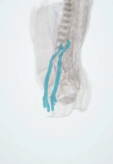
Genitofemoral nerve !
The genitofemoral nerve comes from L1-L2 and runs down the front of the psoas; it carries feeling from the groin and upper inner thigh.
Opens in the Durgical app, no sign-up needed.
What it is
A nerve from L1-L2 that runs down the front of the psoas.
What it does
Carries feeling from the groin and upper inner thigh; at risk in lateral lumbar approaches.
Key facts
- Roots
- L1–L2; emerges on the anterior surface of psoas major
- Moves
- Genital branch: cremaster (efferent limb of cremasteric reflex, L1–L2)
- Feeling from
- Genital branch: anterior scrotum or mons pubis and labium majus; femoral branch: skin over the femoral triangle
Relations
- Course
- Forms from L1–L2 inside psoas, emerges on its front at about L3–L4 and descends under the peritoneum; the genital branch enters the deep inguinal ring, the femoral branch passes behind the inguinal ligament lateral to the femoral artery in the femoral sheath
- In front
- The peritoneum, the ureter and the gonadal vessels, which cross it
- Behind
- The psoas major
How to find it
It cannot be felt. In the retroperitoneum it is seen on the front of the psoas major, usually opposite L3–L4, as one or two thin nerves running down under the peritoneum, behind the ureter; in lateral transpsoas surgery it is at risk where it comes out of the muscle.
Examination
- Cremasteric reflex (L1–L2): stroking the upper inner thigh makes the testis on that side rise; the afferent limb is the femoral branch (and ilioinguinal nerve), the efferent the genital branch; usually absent in testicular torsion
On imaging
Usually not seen on CT or MRI; it is found by its neighbours: on the front of the psoas major, crossed by the ureter, then the genital branch beside the spermatic cord or round ligament in the inguinal canal, where ultrasound can follow it and guide a block. After hernia repair, imaging looks for recurrence or a mesh problem; CT shows a psoas abscess or haematoma that may irritate it.
Clinical notes
- Laparoscopic hernia repair: below the iliopubic tract and lateral to the gonadal vessels lies the 'triangle of pain', with the femoral branch and the lateral femoral cutaneous nerve; tacks placed there can trap them
- Open mesh hernia repair can injure the genital branch on the cord, as well as the ilioinguinal nerve
- Its territory overlaps those of the ilioinguinal and iliohypogastric nerves, so a selective diagnostic block helps to name the nerve responsible
- Renal colic often radiates to the scrotum or labium majus and the upper front of the thigh, partly because the ureter crosses in front of this nerve on the psoas major
Anatomical variants
- Its course varies in over half of people; it can divide early, inside the psoas, so the two branches emerge separately
Related conditions
Test yourself
Think of the answer, then tap the question to see it.
From which roots does the genitofemoral nerve arise, and where does it emerge?
L1–L2; it pierces the psoas major and runs down its anterior surface under the peritoneum.
Which are the afferent and efferent limbs of the cremasteric reflex?
Afferent: the femoral branch of the genitofemoral nerve (and ilioinguinal nerve). Efferent: the genital branch of the genitofemoral nerve to the cremaster.
Where can hernia repair injure the genitofemoral nerve?
The genital branch on the spermatic cord in the inguinal canal (open repair), and the femoral branch in the triangle of pain (laparoscopic tacks).
Educational information, not medical advice. For any symptom, see a health professional.
Its names
- Official Latin (TA2)
- nervus genitofemoralis TA2 6529
- Official Spanish (DeCS)
- Nervio genitofemoral
- English (TA2)
- Genitofemoral nerve
Related
Sources: Terminologia Anatomica (TA2); Anatomy, Abdomen and Pelvis: Genitofemoral Nerve; Anatomía humana (4.ª ed.), tomo 1, cap. 70: Nervios del plexo lumbosacro.
Spotted a mistake?We are always improving for the community. Tell us what is wrong and our team will review it.Report a mistake
Representative model, for educational purposes only.