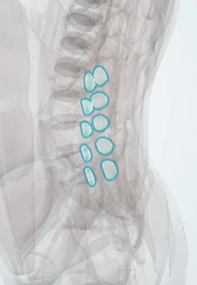
Low-back facet joints !
The lumbar facet joints are pairs of small joints at the back of the lower spine; they block twisting and share the load with the disc.
Official name: Zygapophysial (facet) joints
Opens in the Durgical app, no sign-up needed.
What it is
Pairs of small sliding joints at the back of the spine, one on each side between neighbouring vertebrae, each with cartilage and a capsule.
What it does
Guide and limit movement (lumbar facets block twisting), and share the load with the disc, especially when you lean back.
Key facts
- Where
- Between articular processes from L1–L2 to L5–S1 (zygapophysial joints)
- Function
- Synovial joints; curved near-sagittal facets allow flexion-extension and block rotation; more coronal at L5–S1, resisting forward slip; nerve: medial branches of two dorsal rami
Relations
- In front
- Ligamentum flavum blending with the front of the capsule; just in front, the lateral recess with the traversing root and the foramen with the exiting root
- Behind
- Multifidus, which grips the capsule and the mamillary processes; further out, the erector spinae
- Medial
- Lamina and spinous process; inside the canal, the dural sac with the cauda equina
- Lateral
- Root of the transverse process, where the medial branch runs in a groove under the mamillo-accessory ligament
How to find it
The joints lie deep to the erector spinae, a few centimetres from the midline, at about the level of the gap between two spinous processes. In the paraspinal (Wiltse) approach the facet is the first bony landmark felt between multifidus and longissimus.
Examination
- Facet loading (Kemp) test: standing, the trunk is extended and rotated toward one side; local back pain on that side suggests a facet source, and leg pain below the knee suggests a nerve root. It is not reliable on its own; controlled medial branch blocks are the reference
On imaging
CT shows facet arthropathy best: joint narrowing, osteophytes, subchondral cysts and a vacuum phenomenon. MRI shows effusions and synovial cysts; a large effusion with a slip suggests instability. On the oblique radiograph, a 'collar' on the neck of the Scottie dog marks a pars defect.
Common problems
- Facet arthritis: low back pain that is often worse leaning back or twisting, and stiff in the morning.
- Facet cysts that can press on a nerve root.
- Worn facets allow one vertebra to slip on another (degenerative spondylolisthesis).
How it is diagnosed
There is no single sign. CT or MRI show arthritis; a numbing injection of the nerves to the joint (medial branch block) shows whether the joint is the pain source.
Treatment
- Physical therapy, medicine, facet joint or medial branch injections.
- Radiofrequency ablation: heat turns off the small nerves to the joint for months.
- In a TLIF the facet on one side is removed to reach the disc.
Clinical notes
- Spondylolysis in adolescents who repeat extension and rotation (gymnastics, cricket fast bowling, football); bilateral defects can lead to isthmic spondylolisthesis, usually L5 on S1
- Facet hypertrophy plus ligamentum flavum thickening give the trefoil canal and lateral recess stenosis
- The medial branch groove at the root of the transverse process is the fluoroscopic target for medial branch blocks and radiofrequency neurotomy in facet syndrome
Anatomical variants
- Facet tropism: the left and right facets face different angles, most often at the lower lumbar levels; its link with disc herniation is debated
Memory aid
Scottie dog on the oblique view: eye = pedicle, ear = superior articular process, nose = transverse process, neck = pars, front leg = inferior articular process.
Related conditions
Test yourself
Think of the answer, then tap the question to see it.
Which nerves supply the L4–L5 facet joint?
The L3 and L4 medial branches of the dorsal rami: each lumbar facet joint receives two medial branches, so a block must target two nerves.
Which movement do the lumbar facet joints block, and why?
Rotation: their curved, near-sagittal facets allow flexion-extension but block twisting; at L5–S1 they are more coronal and resist forward slip.
What does a collar on the neck of the Scottie dog on an oblique lumbar radiograph mean?
A pars interarticularis defect (spondylolysis): the neck of the dog is the pars, the stress-fracture site between the upper and lower facets, most often at L5.
Why is degenerative spondylolisthesis most common at L4–L5?
The facets at L4–L5 face sagittally, which favours forward slip once the facets wear.
What is the reference test to confirm that a lumbar facet joint is the pain source?
Controlled medial branch blocks; the facet loading (Kemp) test suggests it but is not reliable on its own.
Educational information, not medical advice. For any symptom, see a health professional.
Its names
- Official Latin (TA2)
- articulationes zygapophysiales TA2 1707
- Official Spanish (DeCS)
- Articulación cigapofisaria DeCS D021801
- English (TA2)
- Zygapophysial (facet) joints
- Also called
- facet joints · juncturae zygapophyseales · zygapophysial joints
Related
Sources: Terminologia Anatomica (TA2); DeCS; Anatomy, Back, Lumbar Spine.
Spotted a mistake?We are always improving for the community. Tell us what is wrong and our team will review it.Report a mistake
Representative model, for educational purposes only.