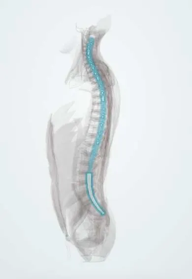
Dura (dural sac) !
The dural sac is a tough membrane tube (the dura) inside the spinal canal, filled with spinal fluid, that holds the spinal cord and nerve roots.
Official name: Spinal dura
Opens in the Durgical app, no sign-up needed.
What it is
A tough tube of membrane (the dura) inside the spinal canal, filled with spinal fluid, holding the spinal cord and nerve roots.
What it does
Protects the cord and nerve roots and keeps them bathed in cerebrospinal fluid.
Key facts
- Where
- Spinal dura from the foramen magnum to about S2; continues as filum terminale externum (coccygeal ligament) to the coccyx
- Attaches
- Edge of foramen magnum; back of C2–C3 bodies; posterior longitudinal ligament (meningovertebral ligaments); coccyx via filum
- Function
- Tough tube enclosing cord, cauda equina and CSF; dural sleeves follow each root to the foramen; epidural space outside it
Relations
- Contents
- Arachnoid and CSF around the spinal cord down to the conus medullaris, then the cauda equina and the filum terminale
- In front
- Anterior epidural space with the internal vertebral venous plexus, then the posterior longitudinal ligament and the discs
- Behind
- Posterior epidural fat, then the ligamentum flavum and the laminae
- Lateral
- Dural root sleeves running into the intervertebral foramina, where they blend with the nerve sheaths; the dorsal root ganglion sits at the foramen
How to find it
In lumbar puncture, after the needle passes the ligamentum flavum, a second, subtler give marks its passage through the dura and arachnoid; with the stylet removed, CSF then drips out. In surgery it appears as a bluish-white tube once the ligamentum flavum and epidural fat are cleared.
On imaging
On T2 MRI bright CSF outlines the sac, the cord and the roots; the sac's cross-sectional area is used to grade central stenosis. CT myelography shows the sac when MRI is not possible. After surgery, a fluid collection behind the sac may be a pseudomeningocele.
Common problems
- Squeezed in spinal stenosis or by a large herniation.
- A tear during surgery (durotomy) can leak spinal fluid; it is usually repaired at once.
How it is diagnosed
MRI shows the sac and how much space it has.
Treatment
- Treated through the cause of the compression. Epidural injections are placed just outside the dura.
Clinical notes
- Lumbar puncture in adults is done below the conus, usually at L3–L4 or L4–L5
- An unrecognised tear during surgery can lead to a pseudomeningocele or a CSF fistula
- Epidural anaesthesia places the drug outside the dura and spinal anaesthesia inside it; accidental dural puncture with an epidural needle is a classic cause of post-dural puncture headache
Anatomical variants
- The lower end of the sac varies, usually between S1 and S3
Development
In newborns the cord and the sac end lower than in adults, and the cord reaches its adult level in the first years of life, so in infants puncture is done at a low lumbar space.
Related conditions
Test yourself
Think of the answer, then tap the question to see it.
Where does the dural sac end, and where does the spinal cord end?
The sac reaches about S2; the cord ends at about L1–L2 (conus medullaris), leaving the lumbar cistern below for lumbar puncture.
What are the features of post-dural puncture headache?
CSF leaks through the dural hole; the headache is worse upright and eases lying down, and is less frequent with thinner, pencil-point needles.
Where is the drug placed in epidural versus spinal anaesthesia relative to the dural sac?
Epidural anaesthesia stays outside the dura; spinal anaesthesia goes inside it, into the CSF.
In which conditions is dural ectasia seen?
Marfan syndrome and neurofibromatosis: a widened dural sac that scallops the back of the vertebral bodies.
Educational information, not medical advice. For any symptom, see a health professional.
Its names
- Official Latin (TA2)
- dura spinalis TA2 5382
- Official Spanish (DeCS)
- Duramadre DeCS D004388
- English (TA2)
- Spinal dura
- Also called
- spinal dura mater · Dura Mater
Related
- Posterior longitudinal ligament
- Ligamentum flavum
- Spinal cord
- Cauda equina
- C4 vertebra
- C5 vertebra
- T1 vertebra
- T2 vertebra
Sources: Terminologia Anatomica (TA2); DeCS; Anatomy, Back, Spinal Meninges; 13.3 Circulation and the Central Nervous System (OpenStax (Rice University), 2022) · adapted, CC BY 4.0.
Spotted a mistake?We are always improving for the community. Tell us what is wrong and our team will review it.Report a mistake
Representative model, for educational purposes only.