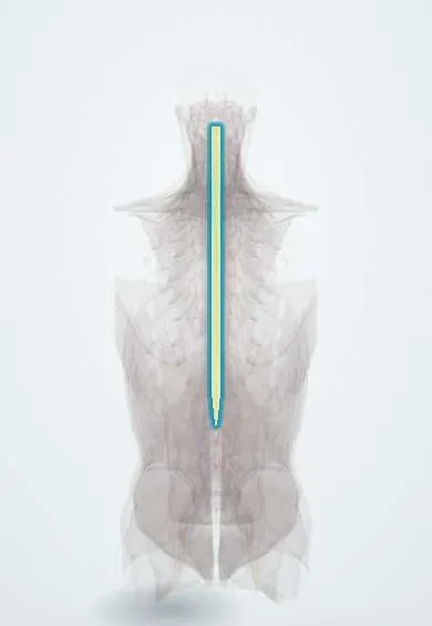
Degenerative cervical myelopathy !
Cervical myelopathy is slow damage to the spinal cord in the neck from a narrowed canal; it causes clumsy hands and an unsteady walk.
Updated 2026-10-29
Structures involved
Tap one to see what it is and open it in 3D.
- Spinal cord
- C5–C6 disc
- Ligamentum flavum
- Posterior longitudinal ligament
- C4–C5 disc
- C6–C7 disc
- Neck facet joints
- C5 vertebra
- C6 vertebra
In the clinical exam
- Babinski sign
- Hoffmann sign and clonus
- Abnormal gaits: hemiparetic, parkinsonian, ataxic, steppage, waddling
- Spinal cord syndromes: complete, Brown-Séquard, central, anterior
In physiology
Why it happens
What it is
Slowly progressive dysfunction of the cervical spinal cord caused by narrowing of the canal from age-related changes of the discs, joints and ligaments. It is the most common cause of spinal cord dysfunction in adults.
Who gets it
Mostly adults over 50, more often men. It is often missed or diagnosed late because early signs are subtle and blamed on age, carpal tunnel syndrome or arthritis.
Causes
- Disc bulging and osteophytes from cervical spondylosis (the most common cause)
- Thickening or buckling of the ligamentum flavum
- Ossification of the posterior longitudinal ligament (OPLL)
- Degenerative slip of one vertebra on another (spondylolisthesis) or instability
Risk factors
- Older age
- Congenitally narrow cervical canal
- Manual labour or contact sports with repeated neck loading
- Previous neck surgery or fusion at a neighbouring level
- Minor trauma (hyperextension) in a narrowed canal, which can cause acute worsening
Pathophysiology
The narrowed canal compresses the cord, both statically and dynamically with neck flexion and extension. Repeated mechanical stress and reduced blood flow cause ischaemia, demyelination and loss of neurons in the grey and white matter. Upper motor neuron signs appear below the level, often with lower motor neuron signs at the level.
Classification
- Severity by the modified Japanese Orthopaedic Association scale (mJOA, 18 points): mild, moderate or severe
- Commonly used cut-offs: mild 15–17, moderate 12–14, severe 11 or less
- By the main compressing structure: disc-osteophyte, ligamentum flavum, OPLL, or a mix
Symptoms and signs
- Clumsy hands: trouble with buttons, writing or picking up coins; numb hands without a single nerve pattern
- Unsteady, wide-based gait and poor balance, especially in the dark
- Hoffmann sign: flicking the middle fingernail makes the thumb and index finger flex
- Hyperreflexia, clonus, Babinski sign and leg spasticity
- Neck stiffness or pain, which may be absent
- Lhermitte sign: electric shock down the spine on neck flexion
- Bladder urgency or incontinence, usually late
Diagnosis
- Clinical
- History of hand clumsiness and gait change with upper motor neuron signs (Hoffmann sign, hyperreflexia, Babinski, inverted brachioradialis reflex, clonus) and Romberg or tandem-gait testing. The mJOA score grades severity and follows progression.
- Tests
- Blood tests for look-alikes when the picture is unclear: vitamin B12, copper and others. Electromyography and nerve conduction help separate it from peripheral neuropathy or motor neuron disease.
- Imaging
- MRI of the cervical spine is the study of choice: it shows cord compression and signal change in the cord. X-rays (with flexion-extension views) show alignment and instability; CT shows OPLL and osteophytes.
Differential diagnosis
- Carpal tunnel syndrome or other peripheral nerve entrapment
- Cervical radiculopathy
- Amyotrophic lateral sclerosis
- Multiple sclerosis or transverse myelitis
- Subacute combined degeneration (vitamin B12 or copper deficiency)
- Spinal cord tumour
Goal of treatment
Stop neurological decline and, where possible, recover function by decompressing the cord in the right patients, while protecting the cord from further injury.
Treatment
- Follow-up
- Mild cases may be followed closely with repeated neurological exams and mJOA scores; surgery is offered if function worsens. Neck manipulation is avoided.
- Rehabilitation
- Supervised physiotherapy for mild disease and after surgery: balance, gait and hand training, with fall prevention.
- Surgical
- Anterior decompression for compression at one to three levels from the front (discs, osteophytes): anterior cervical discectomy and fusion or corpectomy.
- Surgical
- Posterior decompression for multilevel disease or OPLL: laminectomy with fusion or laminoplasty. Surgery is generally recommended for moderate and severe myelopathy.
Complications
- Progressive weakness, falls and loss of independence
- Acute spinal cord injury (central cord syndrome) after minor trauma
- Neurogenic bladder
- Surgical complications: C5 nerve root palsy, dysphagia, adjacent-segment disease, infection
Prognosis
The natural course is variable but often stepwise decline. Surgery reliably halts progression and usually improves function; outcomes are better when it is done before severe or long-standing deficits develop.
Test yourself
Think of the answer, then tap the question to see it.
What is the most common cause of spinal cord dysfunction in adults?
Degenerative cervical myelopathy, from age-related narrowing of the cervical canal.
How is the Hoffmann sign elicited and what does it suggest?
Flicking the nail of the middle finger makes the thumb and index finger flex. It suggests an upper motor neuron lesion such as cervical cord compression.
Which scale grades the severity of cervical myelopathy?
The modified Japanese Orthopaedic Association scale (mJOA), out of 18 points: lower scores mean worse function.
What is the imaging study of choice in suspected cervical myelopathy?
MRI of the cervical spine, which shows cord compression and signal change within the cord.
When is surgery generally recommended for degenerative cervical myelopathy?
For moderate or severe myelopathy, and for mild disease that worsens or does not respond to supervised nonoperative care.
Related conditions
- Cervical Spondylosis
- Cervical radiculopathy
- Spinal cord injury
- Carpal tunnel syndrome
- Multiple sclerosis
Educational information, not medical advice. For any symptom, see a health professional.
Sources: Cervical Myelopathy (StatPearls (NCBI Bookshelf), 2025).
Spotted a mistake?We are always improving for the community. Tell us what is wrong and our team will review it.Report a mistake
Representative model, for educational purposes only.