Conditions · Ophthalmology and ENT
Juvenile nasopharyngeal angiofibroma !
A rare, benign but very vascular tumour at the back of the nose in adolescent boys. It causes a blocked nose and heavy, repeated nosebleeds.
Updated 2026-11-01
Structures involved
Tap one to see what it is and open it in 3D.
- Nasal cavity
- Pharynx
- Maxillary artery
- Sphenoid sinus
- Sphenoid bone
- Maxillary sinus
- External carotid artery
- Auditory tube
- Cavernous sinus
- Inferior nasal conchae
- Nasal septum
Why it happens
- Benign vs malignant tumours: the comparison table
- Testosterone and its actions
- Blood vessels: artery, vein and capillary layers
What it is
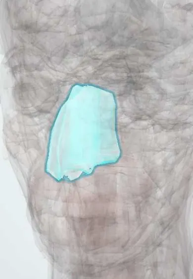
A benign, highly vascular fibrous tumour that arises near the sphenopalatine foramen on the posterolateral wall of the nasal cavity and grows into the nasopharynx. Although it does not metastasise, it is locally aggressive and can erode bone and spread into the sinuses, the pterygopalatine and infratemporal fossae, the orbit and the skull base.
Who gets it
Rare: about 0.5% of head and neck tumours. It occurs almost only in males, mostly between about 10 and 25 years of age. Any adolescent boy with recurrent one-sided nosebleeds and nasal blockage should raise suspicion.
Causes
- Exact cause unknown
- Hormonal influence is suspected (androgen-sensitive tissue), explaining the male adolescent predominance
- Possible origin from vascular remnants near the sphenopalatine foramen
- Higher frequency reported in familial adenomatous polyposis
Risk factors
- Male sex
- Adolescence (puberty)
- Family history of familial adenomatous polyposis
Pathophysiology
The tumour is made of fibrous stroma with many thin-walled vessels that lack a normal muscular layer, so they do not contract and bleed heavily. It is fed mainly by branches of the maxillary artery (from the external carotid), especially the sphenopalatine artery, and in large tumours also by internal carotid branches. As it grows it blocks the choana and the auditory tube, bows the posterior wall of the maxillary sinus forward, widens the pterygopalatine fossa and can reach the orbit, cavernous sinus and middle cranial fossa.
Classification
- Staged by extent on imaging (Fisch, Radkowski or UPMC systems)
- Confined to nasal cavity and nasopharynx without bone destruction
- Spread into sinuses or pterygopalatine fossa with bone erosion
- Extension to infratemporal fossa, orbit or skull base
- Intracranial extension, cavernous sinus or optic chiasm involvement
Symptoms and signs
- Progressive one-sided nasal obstruction
- Recurrent, heavy, one-sided nosebleeds that are hard to stop
- Nasal discharge and hyponasal voice
- Ear fullness or hearing loss from blocked auditory tube (middle ear effusion)
- Swelling of the cheek, proptosis or double vision in advanced cases
- Headache
Diagnosis
- Clinical
- Typical history in an adolescent male. Nasal endoscopy shows a smooth, lobulated, reddish-purple mass in the posterior nasal cavity and nasopharynx.
- Tests
- Office biopsy is generally avoided because of the risk of life-threatening bleeding; the diagnosis is usually made on characteristic imaging and confirmed on the resected specimen. Blood count and cross-matching before surgery because of expected blood loss.
- Imaging
- Contrast CT shows an avidly enhancing mass centred on the sphenopalatine foramen, widening of the pterygopalatine fossa and anterior bowing of the posterior maxillary wall (Holman–Miller sign). MRI defines soft-tissue and intracranial extent. Angiography maps the feeding vessels and allows preoperative embolisation.
Differential diagnosis
- Antrochoanal or nasal polyp
- Nasopharyngeal carcinoma
- Adenoid hypertrophy
- Rhabdomyosarcoma or lymphoma in children and adolescents
- Other causes of recurrent epistaxis (anterior septal bleeding, coagulopathy)
- Inverted papilloma
Goal of treatment
Complete removal of the tumour with control of bleeding, preservation of surrounding structures and close follow-up for recurrence.
Treatment
- Acute
- Massive epistaxis: airway protection, nasal packing, intravenous fluids and transfusion as needed, and urgent ENT and interventional radiology involvement.
- Procedure
- Preoperative angiographic embolisation of the feeding branches of the maxillary artery, usually one to a few days before surgery, to reduce intraoperative blood loss.
- Surgical
- Surgical excision is the treatment of choice: endoscopic endonasal resection for most tumours, with open or combined approaches for extensive disease.
- Medical
- Radiotherapy is reserved for unresectable, intracranial or residual disease; hormonal (anti-androgen) therapy has been studied but is not standard.
- Follow-up
- Postoperative care: monitoring for bleeding, airway and haemoglobin, care of nasal packing; then serial endoscopy and MRI to detect recurrence, which is most frequent in the first years.
Complications
- Life-threatening haemorrhage, spontaneous or during biopsy or surgery
- Recurrence or residual tumour
- Intracranial and orbital extension with visual loss
- Middle ear effusion and conductive hearing loss
- Long-term effects of radiotherapy on growth and the skull base in young patients
Prognosis
Survival is excellent with treatment. Recurrence is reported in a substantial minority, mostly with extensive or skull-base disease and incomplete resection; follow-up imaging is needed for years. Some tumours slowly regress after adolescence.
Test yourself
Think of the answer, then tap the question to see it.
What is the classic patient with juvenile nasopharyngeal angiofibroma?
An adolescent male with progressive unilateral nasal obstruction and recurrent, heavy unilateral epistaxis.
Which artery mainly supplies the tumour?
The maxillary artery (a branch of the external carotid), mainly through the sphenopalatine artery.
Why is office biopsy usually avoided?
The tumour is highly vascular and its vessels lack muscle in their walls, so biopsy can cause severe, life-threatening bleeding.
What is the Holman–Miller sign?
Anterior bowing of the posterior wall of the maxillary sinus on CT, caused by tumour in the pterygopalatine fossa.
What is done before surgical resection to reduce bleeding?
Angiography with embolisation of the feeding vessels.
Related conditions
- Epistaxis (nosebleed)
- Nasopharyngeal carcinoma
- Chronic rhinosinusitis and nasal polyps
- Acute rhinosinusitis
- Allergic rhinitis
- Otitis media with effusion and chronic suppurative otitis media
Educational information, not medical advice. For any symptom, see a health professional.
Sources: Juvenile Nasopharyngeal Angiofibroma (JNA) (Cleveland Clinic Health Library, 2025); Juvenile nasopharyngeal angiofibroma (Journal of Oral and Maxillofacial Pathology, 2016).
Spotted a mistake?We are always improving for the community. Tell us what is wrong and our team will review it.Report a mistake
Representative model, for educational purposes only.