Nervous system · Head and neck
Hippocampus !
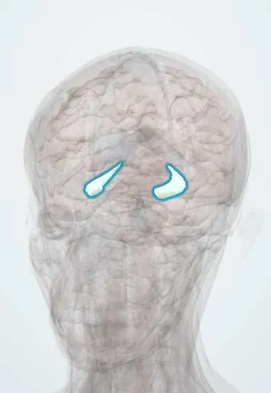
The hippocampus is a curved, seahorse-shaped ridge deep in the temporal lobe; it forms new memories and is affected early in Alzheimer disease.
Opens in the Durgical app, no sign-up needed.
What it is
A curved seahorse-shaped ridge in the floor of the temporal horn.
What it does
Forms new memories and helps navigation; affected early in Alzheimer disease.
Key facts
- Where
- Floor of the temporal horn of the lateral ventricle
- Blood supply
- PCA, anterior choroidal artery
- Function
- Forms new memories (affected early in Alzheimer); spatial navigation
Relations
- Contents
- Dentate gyrus, cornu ammonis (CA1–CA4) and subiculum; the main circuit runs entorhinal cortex, dentate gyrus, CA3, CA1, subiculum
- Above
- Temporal horn of the lateral ventricles, whose floor it forms, with the choroid plexus; the fimbria on its upper edge leads to the fornix
- In front
- Amygdala, separated from it by the tip of the temporal horn
- Medial
- Ambient cistern, the midbrain and the posterior cerebral artery
- Behind
- Tail curving up behind the thalamus, where the fimbria becomes the crus of the fornix under the splenium
- Below
- Parahippocampal gyrus (subiculum), continuous with it below the hippocampal sulcus, which separates it from the dentate gyrus; lateral to it in the ventricle floor, the collateral eminence over the collateral sulcus
How to find it
Seen on coronal MRI angled perpendicular to its long axis: a curled grey structure in the floor and medial wall of the temporal horn, with head, body and tail from front to back. The head shows small ridges (digitations), which are lost in sclerosis.
Examination
- Three-word recall after a short delay: the words are repeated at once but lost minutes later when consolidation of new memories fails, as after bilateral hippocampal damage
- Intracarotid amobarbital (Wada) test before epilepsy surgery: shows whether the other side can support memory and which side holds language; functional MRI now often replaces it
On imaging
Mesial temporal sclerosis: small hippocampus with high T2/FLAIR signal and loss of internal structure. Alzheimer disease: volume loss with a widened temporal horn and choroidal fissure, graded on coronal T1 with visual scales. Transient global amnesia may show tiny diffusion dots in CA1.
Common problems
- Alzheimer disease shrinks it early, so new memories fade first.
- A common source of seizures (temporal lobe epilepsy).
How it is diagnosed
Memory tests; MRI shows its size.
Treatment
- Medicines and support for dementia; seizure medicines; epilepsy surgery in selected cases.
Clinical notes
- Selective amygdalohippocampectomy or anterior temporal lobectomy can stop seizures in selected people with mesial temporal sclerosis
- Autoimmune (limbic) encephalitis and herpes simplex encephalitis inflame the hippocampi, with memory loss, seizures and behaviour change
Anatomical variants
- Incomplete hippocampal inversion: a rounder, more upright hippocampus, more often on the left; usually a normal finding, not to be mistaken for sclerosis (some texts)
Related conditions
- Sanfilippo syndrome (mucopolysaccharidosis type III)
- Epilepsy and seizures
- Alzheimer disease
- Isoniazid toxicity
- Cannabis toxicity and cannabinoid hyperemesis syndrome
- Dementia (major neurocognitive disorder)
- Mild cognitive impairment
- Rabies
- Viral encephalitis (herpes simplex encephalitis)
- Status epilepticus
- Autoimmune encephalitis (including anti-NMDA receptor encephalitis)
- Febrile seizures
- Major depressive disorder
- Schizophrenia
- Generalized anxiety disorder
- Post-traumatic stress disorder (PTSD) and acute stress disorder
- Cannabis use disorder
- Dissociative disorders
Test yourself
Think of the answer, then tap the question to see it.
What does bilateral damage to the hippocampus cause?
Anterograde amnesia, as the hippocampus consolidates new declarative memories; older memories and skills are largely spared.
Which hippocampal sector is most sensitive to lack of oxygen?
CA1 (Sommer sector); cardiac arrest can leave lasting memory loss.
What is mesial temporal sclerosis, and how does it appear on MRI?
A shrunken, scarred hippocampus, the most common cause of drug-resistant temporal lobe epilepsy; MRI shows a small hippocampus with high T2/FLAIR signal.
Where does the hippocampus lie?
In the floor of the temporal horn of the lateral ventricle, with the amygdala in front; the fimbria on its upper edge leads to the fornix.
Educational information, not medical advice. For any symptom, see a health professional.
Its names
- Official Latin (TA2)
- hippocampus TA2 5518
- Official Spanish (DeCS)
- Hipocampo DeCS D006624
- English (TA2)
- Hippocampus
- Also called
- Ammon's horn · hippocampal formation
Related
Sources: Terminologia Anatomica (TA2); DeCS; Neuroanatomy, Hippocampus; 16.2 The Mental Status Exam (OpenStax (Rice University), 2022) · adapted, CC BY 4.0; Tratado de anatomía humana (9.ª ed.), tomo II, Libro V, cap. VI: Cerebro (configuración exterior e interior, ventrículos, formaciones coroideas, epífisis).
Spotted a mistake?We are always improving for the community. Tell us what is wrong and our team will review it.Report a mistake
Representative model, for educational purposes only.