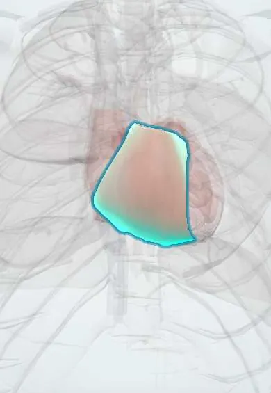
Arrhythmogenic right ventricular cardiomyopathy !
An inherited heart muscle disease in which fat and scar replace the right ventricle's muscle, causing dangerous rhythms in young people.
Updated 2026-11-01
Structures involved
Tap one to see what it is and open it in 3D.
- Right ventricle
- Pulmonary trunk
- Tricuspid anterior leaflet
- Tricuspid septal leaflet
- Tricuspid posterior leaflet
- Interventricular septum
- Left ventricle
- Bundle branches
Why it happens
- Mendelian inheritance: autosomal dominant, recessive and X-linked
- Fibrosis in organs: TGF-β and the myofibroblast
- How arrhythmias arise
- Cell junctions: tight, adherens, desmosomes, gap, hemidesmosomes
- Cardiac muscle: intercalated discs
What it is
Arrhythmogenic right ventricular cardiomyopathy (ARVC) is an inherited disease of the desmosomes, the junctions that hold heart muscle cells together. Myocytes are progressively replaced by fibrous and fatty tissue, mainly in the right ventricle, causing ventricular arrhythmias, right ventricular dilation and, later, heart failure. It is a leading cause of sudden cardiac death in young people and athletes.
Who gets it
Estimated to affect about 1 in 1,000 to 1 in 5,000 people and probably underdiagnosed. It usually presents between adolescence and the fourth decade, more often and more severely in men. Up to half of cases run in families. There are no specific Colombian data.
Causes
- Pathogenic variants in desmosome genes: PKP2 (plakophilin-2, the most common), DSP, DSG2, DSC2, JUP
- Less often non-desmosomal genes (for example TMEM43, PLN, DES, LMNA)
- Mostly autosomal dominant with variable penetrance; rare recessive forms with skin and hair findings (Naxos disease, Carvajal syndrome)
- Endurance exercise accelerates disease expression in carriers
Risk factors
- Family history of ARVC or of unexplained sudden death in a young relative
- Carrying a pathogenic desmosomal variant
- Male sex
- High-intensity and endurance sport
Pathophysiology
Defective desmosomes cannot withstand the mechanical stress of contraction, so cell junctions detach and myocytes die. Repair replaces them with fibrofatty tissue, beginning under the epicardium and in the so-called triangle of dysplasia (inflow, outflow and apex of the right ventricle). Islands of surviving muscle within scar create re-entry circuits that cause monomorphic ventricular tachycardia with a left bundle branch block pattern. Delayed conduction across diseased tissue produces epsilon waves. Progression causes right ventricular dilation and dysfunction, and the left ventricle may also be involved.
Classification
- Classic right-dominant form
- Biventricular form
- Left-dominant form (arrhythmogenic left ventricular cardiomyopathy)
- Diagnostic categories of the Task Force criteria: definite, borderline, possible
Symptoms and signs
- Palpitations
- Light-headedness or syncope, often during exercise
- Cardiac arrest as the first sign in some young patients
- Later: dyspnoea, fatigue, leg and abdominal swelling from right heart failure
- Many relatives carry the gene without symptoms
Diagnosis
- Clinical
- Diagnosis combines several criteria (Task Force criteria): structural and functional abnormalities of the right ventricle, tissue characterisation, repolarisation and depolarisation abnormalities on the ECG, arrhythmias, and family history or genetics. Suspect it in a young person with exercise-related palpitations or syncope.
- Tests
- 12-lead ECG: T-wave inversion in V1 to V3, epsilon waves, prolonged terminal activation of QRS. Holter monitoring shows frequent premature ventricular contractions or ventricular tachycardia with left bundle branch block morphology. Signal-averaged ECG detects late potentials. Genetic testing of the patient and cascade testing of first-degree relatives.
- Imaging
- Echocardiography shows right ventricular dilation, reduced function and regional wall motion abnormalities or aneurysms. Cardiac magnetic resonance is the best imaging test, adding fibrofatty replacement and late gadolinium enhancement. Endomyocardial biopsy is rarely needed.
Differential diagnosis
- Idiopathic right ventricular outflow tract tachycardia
- Brugada syndrome
- Athlete's heart
- Cardiac sarcoidosis and myocarditis
- Dilated cardiomyopathy
- Right ventricular volume overload (atrial septal defect, pulmonary hypertension)
Goal of treatment
Prevent sudden cardiac death, control arrhythmias, slow progression and screen family members.
Treatment
- Lifestyle
- Restriction of competitive and endurance sport for patients and gene-positive relatives, the main disease-modifying measure.
- Procedure
- Implantable cardioverter-defibrillator for survivors of cardiac arrest, sustained ventricular tachycardia, unexplained syncope or other high-risk features.
- Medical
- Beta-blockers for all affected patients; amiodarone or sotalol as antiarrhythmics when arrhythmias persist.
- Procedure
- Catheter ablation (endocardial and epicardial) for recurrent ventricular tachycardia despite drugs.
- Medical
- Standard heart failure therapy for ventricular dysfunction; heart transplantation in refractory heart failure or arrhythmias.
- Follow-up
- Nursing and follow-up: defibrillator education, telemetry after shocks, regular ECG and imaging, genetic counselling and periodic screening of relatives.
Complications
- Sudden cardiac death (often during exercise)
- Sustained ventricular tachycardia and electrical storm
- Right or biventricular heart failure
- Thromboembolism in a dilated, poorly contracting ventricle
- Inappropriate defibrillator shocks and psychological impact
Prognosis
With early diagnosis, exercise restriction and a defibrillator when indicated, long-term survival is good. Risk is highest in young patients with syncope, sustained arrhythmias, extensive disease or left ventricular involvement. Disease tends to progress over decades.
Prevention
- Cascade genetic and clinical screening of first-degree relatives
- Pre-participation screening of athletes with ECG where available
- Avoidance of endurance and competitive sport in carriers
Test yourself
Think of the answer, then tap the question to see it.
Which cell structure is defective in ARVC?
The desmosome (for example plakophilin-2).
What is the characteristic ECG finding?
Epsilon waves and T-wave inversion in V1 to V3.
What QRS morphology does ventricular tachycardia from the right ventricle show?
A left bundle branch block pattern.
Which lifestyle measure is essential?
Avoiding competitive and endurance sport.
What is the best imaging test for diagnosis?
Cardiac magnetic resonance.
Related conditions
- Ventricular tachycardia
- Cardiac arrest (VF, pulseless VT, asystole, PEA)
- Syncope (vasovagal, orthostatic hypotension, cardiac)
- Brugada syndrome
- Premature atrial and ventricular contractions
- Dilated cardiomyopathy
- Hypertrophic cardiomyopathy
- Myocarditis
- Sarcoidosis
- Heart failure
- Atrial septal defect
- Pulmonary hypertension and cor pulmonale
Educational information, not medical advice. For any symptom, see a health professional.
Sources: Arrhythmogenic right ventricular cardiomyopathy: MedlinePlus Genetics (MedlinePlus Genetics, US National Library of Medicine, 2026); Cardiomyopathy - Types | NHLBI, NIH (National Heart, Lung, and Blood Institute (NHLBI), NIH, 2026).
Spotted a mistake?We are always improving for the community. Tell us what is wrong and our team will review it.Report a mistake
Representative model, for educational purposes only.