Nervous system · Head and neck
Caudate nucleus !
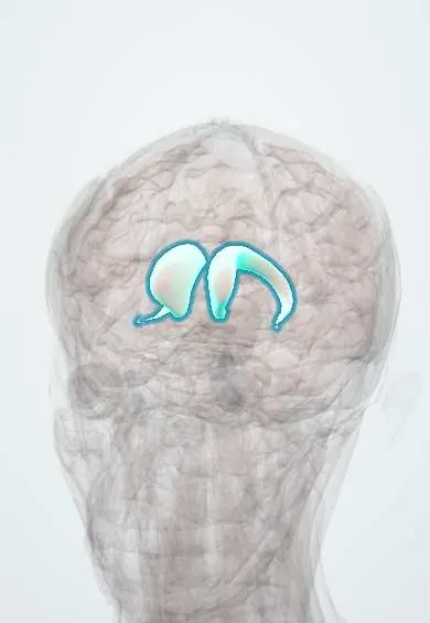
The caudate nucleus is a C-shaped mass deep in the brain, part of the basal ganglia; it helps start and smooth movement and shrinks in Huntington disease.
Opens in the Durgical app, no sign-up needed.
What it is
A C-shaped deep nucleus curving along the lateral ventricle.
What it does
Part of the basal ganglia: helps start and smooth movement and habits; it shrinks in Huntington disease.
Key facts
- Where
- Curves along the lateral ventricle (head, body, tail)
- Blood supply
- Lenticulostriate arteries (MCA), recurrent artery of Heubner (ACA); anterior choroidal artery (body and tail)
- Function
- Basal ganglia: movement, habits, cognition (atrophies in Huntington)
Relations
- Course
- Head in the lateral wall of the frontal horn of the lateral ventricles; body along the floor of the ventricle's body; the tail arches back and down into the roof of the temporal horn and ends near the amygdala
- Medial
- In front, the frontal horn with the septum pellucidum beyond it; further back the thalamus, separated by a groove that holds the stria terminalis and the thalamostriate vein
- Lateral
- The anterior limb of the internal capsule separates the head from the putamen; grey bridges cross the capsule between them, the stripes that gave the striatum its name
- Below
- Below and in front, the head merges with the putamen at the nucleus accumbens (ventral striatum), just above the anterior perforated substance
How to find it
On axial and coronal CT or MRI its head bulges into the lateral wall of each frontal horn, with the anterior limb of the internal capsule between it and the putamen; body and tail thin out as they follow the ventricle back and into the temporal lobe. Opened from above, the lateral ventricle shows it in its floor and lateral wall.
Examination
- Motor impersistence: the person cannot keep the tongue out or hold a steady grip (the grip squeezes and relaxes, "milkmaid grip"), a sign of chorea as in Huntington disease
- Saccades: slow, delayed voluntary saccades, sometimes started with a head thrust or a blink, are early signs in Huntington disease
On imaging
On axial CT or MRI the head is the grey bulge forming the lateral wall of each frontal horn. In Huntington disease the gap between the two caudate heads widens as the frontal horns enlarge. On cranial ultrasound in preterm babies, the groove between the caudate and the thalamus (caudothalamic groove) is the usual site of germinal matrix haemorrhage.
Clinical notes
- Hypertensive bleeding in the caudate often breaks into the lateral ventricle and can mimic a subarachnoid haemorrhage, with sudden headache and neck stiffness
- The recurrent artery of Heubner, which feeds the caudate head and the anterior limb of the internal capsule, can be injured when an aneurysm of the anterior communicating artery is clipped; the opposite face and arm weaken
Development
It grows from the lateral ganglionic eminence in the floor of the embryonic lateral ventricle; its C shape follows the ventricle as the hemisphere curls back and down to form the temporal lobe.
Memory aid
Striatum = caudate + putamen (the 'striped' pair); lentiform nucleus = putamen + globus pallidus (the 'lens').
Related conditions
- Huntington disease
- Lesch-Nyhan syndrome
- Leigh syndrome
- Parkinson disease
- Perinatal asphyxia and hypoxic-ischaemic encephalopathy
- Cerebral palsy
- Intraventricular haemorrhage of prematurity
- Schizophrenia
- Obsessive-compulsive disorder (OCD)
- Attention-deficit/hyperactivity disorder (ADHD)
- Stimulant use disorder (cocaine, amphetamines)
- Antipsychotic-induced movement disorders (dystonia, akathisia, parkinsonism, tardive dyskinesia)
- Tourette syndrome and tic disorders
- Body dysmorphic disorder, hoarding disorder and trichotillomania
- Gambling disorder
Test yourself
Think of the answer, then tap the question to see it.
How does Huntington disease change the caudate nucleus on imaging?
The caudate head wastes away, so the frontal horns lose their inward bulge and look squared-off («boxcar»).
Which structures form the striatum?
The caudate nucleus and the putamen; the striatum is the input station of the basal ganglia, receiving excitatory fibres from the cortex and dopamine from the substantia nigra.
Why do caudate lesions change behaviour more than movement?
The caudate nucleus is wired mainly to the prefrontal cortex, so lesions cause apathy, loss of initiative or disinhibition.
Which artery feeding the caudate head can be injured when an anterior communicating artery aneurysm is clipped?
The recurrent artery of Heubner, which also feeds the anterior limb of the internal capsule; the opposite face and arm weaken.
Educational information, not medical advice. For any symptom, see a health professional.
Its names
- Official Latin (TA2)
- nucleus caudatus TA2 5561
- Official Spanish (DeCS)
- Núcleo Caudado DeCS D002421
- English (TA2)
- Caudate nucleus
Related
Sources: Terminologia Anatomica (TA2); DeCS; Neuroanatomy, Nucleus Caudate; 13.2 The Central Nervous System (OpenStax (Rice University), 2022) · adapted, CC BY 4.0.
Spotted a mistake?We are always improving for the community. Tell us what is wrong and our team will review it.Report a mistake
Representative model, for educational purposes only.