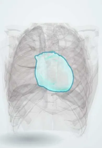
Constrictive pericarditis !
The sac around the heart becomes thick and scarred like a rigid shell, so the heart cannot fill. It causes swelling and can be cured by surgery.
Updated 2026-11-01
Structures involved
Tap one to see what it is and open it in 3D.
- Pericardium
- Right ventricle
- Left ventricle
- Right atrium
- Interventricular septum
- Inferior vena cava
- Superior vena cava
In the clinical exam
- Jugular venous pressure and the venous pulse
- Hepatojugular (abdominojugular) reflux
- Cardiac tamponade: Beck triad and pulsus paradoxus
Why it happens
- Fibrosis in organs: TGF-β and the myofibroblast
- Outcomes of acute inflammation: resolution, abscess, scar or chronic inflammation
- Patterns of acute inflammation: serous, fibrinous, purulent, abscess, ulcer
- The cardiac cycle
- Central venous pressure and the jugular pulse
What it is
Constrictive pericarditis is a chronic condition in which a thickened, fibrotic and often calcified pericardium encases the heart and limits diastolic filling. It produces mainly right-sided heart failure with a normal-sized heart. It is important because it is potentially curable by surgical removal of the pericardium.
Who gets it
Uncommon. In high-income countries most cases follow cardiac surgery, radiation or idiopathic/viral pericarditis. In regions where tuberculosis is frequent, including parts of Latin America and Colombia, tuberculous pericarditis remains a leading cause, and constriction develops in a substantial proportion of those patients.
Causes
- Idiopathic or viral pericarditis
- Previous cardiac surgery
- Mediastinal radiation therapy (for example for lymphoma or breast cancer)
- Tuberculosis (the main cause in endemic regions)
- Connective tissue disease, purulent pericarditis, uraemia, malignancy, trauma
Risk factors
- Previous acute or recurrent pericarditis, especially tuberculous or purulent
- Previous heart surgery or chest radiation
- HIV infection with tuberculosis
- Chronic kidney disease and autoimmune disease
Pathophysiology
The rigid pericardium fixes the total cardiac volume. Early diastolic filling is rapid and then stops abruptly when the heart meets the shell, producing a pericardial knock and a «dip and plateau» ventricular pressure. Intrathoracic pressure changes are not transmitted to the heart chambers, so inspiration fails to increase venous return (Kussmaul sign) and the ventricles compete for space: right ventricular filling rises as left ventricular filling falls (ventricular interdependence). Diastolic pressures equalise in all four chambers and systemic venous congestion dominates.
Classification
- Chronic constrictive pericarditis
- Transient constriction (resolves with anti-inflammatory therapy, often after recent pericarditis)
- Effusive-constrictive pericarditis (effusion plus constricting visceral pericardium)
Symptoms and signs
- Slowly progressive exertional dyspnoea, fatigue and weakness
- Leg oedema, abdominal swelling (ascites) and an enlarged liver
- Markedly raised jugular venous pressure that rises on inspiration (Kussmaul sign) with a sharp y descent
- Pericardial knock (early diastolic sound)
- Weight loss and muscle wasting in long-standing disease
Diagnosis
- Clinical
- Suspect it in right-sided heart failure with ascites and oedema but no lung disease or valve disease and a normal-sized heart, especially after pericarditis, surgery, radiation or tuberculosis. The jugular venous pressure is high, Kussmaul sign is present and a pericardial knock may be heard. Pulsus paradoxus is less common than in tamponade.
- Tests
- BNP is typically only mildly raised (lower than expected for the degree of congestion), a clue against restrictive cardiomyopathy. ECG may show low voltage, non-specific T-wave changes or atrial fibrillation. Tuberculosis work-up where relevant. Cardiac catheterisation: equal diastolic pressures in all chambers, dip-and-plateau pattern and discordant respiratory changes in left and right ventricular systolic pressures.
- Imaging
- Chest X-ray may show pericardial calcification (best seen on the lateral view). Echocardiography shows septal bounce, respirophasic septal shift, marked respiratory variation in mitral inflow, preserved or increased medial tissue Doppler velocity (annulus reversus) and a dilated inferior vena cava. CT or cardiac MRI demonstrate pericardial thickening, calcification and active inflammation.
Differential diagnosis
- Restrictive cardiomyopathy
- Cardiac tamponade
- Right heart failure from pulmonary hypertension or tricuspid valve disease
- Cirrhosis with ascites
- Nephrotic syndrome
Goal of treatment
Relieve venous congestion, treat any active cause such as tuberculosis, and remove the constricting pericardium in chronic disease before advanced myocardial atrophy or liver damage occur.
Treatment
- Medical
- Anti-inflammatory therapy (NSAIDs, colchicine, sometimes corticosteroids) for transient or inflammatory constriction, which may reverse without surgery.
- Medical
- Antituberculous treatment for tuberculous pericarditis, which lowers the risk of progression to constriction.
- Medical
- Diuretics and a low-salt diet to relieve congestion as a bridge or for patients who cannot undergo surgery.
- Surgical
- Pericardiectomy (surgical removal of the pericardium) is the definitive treatment for chronic constriction.
- Follow-up
- Daily weight, fluid balance, abdominal girth, jugular venous pressure, kidney function and electrolytes; postoperative monitoring for low cardiac output and arrhythmias.
Complications
- Chronic right-sided heart failure, ascites and protein-losing enteropathy
- Congestive hepatopathy and cardiac cirrhosis
- Atrial fibrillation
- Low cardiac output after pericardiectomy, particularly in radiation-induced disease
Prognosis
Pericardiectomy relieves symptoms in most patients, with the best results in idiopathic cases and the worst after radiation, where the myocardium itself is often damaged. Untreated chronic constriction progresses steadily.
Prevention
- Early diagnosis and full treatment of tuberculosis, including tuberculous pericarditis
- Adequate treatment of acute pericarditis and drainage of purulent pericarditis
- Cardiac-sparing radiation techniques
Test yourself
Think of the answer, then tap the question to see it.
What is the classic physical sign of a rise in jugular venous pressure on inspiration?
Kussmaul sign.
Which early diastolic sound is typical of constrictive pericarditis?
The pericardial knock.
Which infectious cause is most important in developing countries?
Tuberculosis.
Which catheterisation finding is shared with restrictive cardiomyopathy?
The dip-and-plateau (square root) diastolic pressure pattern.
What is the definitive treatment for chronic constriction?
Surgical pericardiectomy.
Related conditions
- Acute pericarditis
- Cardiac tamponade
- Restrictive cardiomyopathy (amyloid, sarcoid, haemochromatosis)
- Tuberculosis
- Heart failure
- Cirrhosis
- Atrial fibrillation
Educational information, not medical advice. For any symptom, see a health professional.
Sources: Pericarditis - constrictive (MedlinePlus, US National Library of Medicine, 2026).
Spotted a mistake?We are always improving for the community. Tell us what is wrong and our team will review it.Report a mistake
Representative model, for educational purposes only.