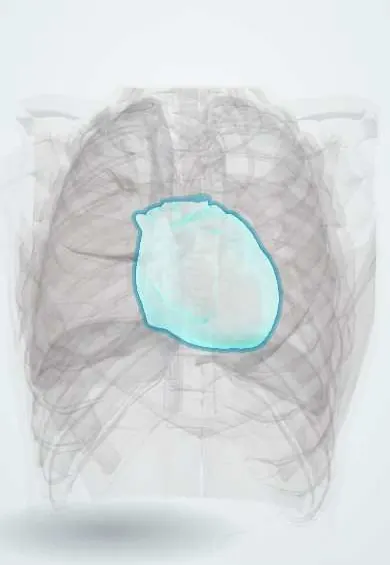
Cardiac tamponade !
Cardiac tamponade is life-threatening pressure on the heart from fluid or blood in the sac around it, which stops the heart filling properly.
Updated 2026-10-29
Structures involved
Tap one to see what it is and open it in 3D.
- Pericardium
- Right atrium
- Right ventricle
- Superior vena cava
- Inferior vena cava
- Left ventricle
- Ascending aorta
- Sternum
In the clinical exam
- Jugular venous pressure and the venous pulse
- Abnormal pulses: paradoxus, alternans, parvus et tardus, bounding
- Cardiac tamponade: Beck triad and pulsus paradoxus
- Shock: the clinical signs and the types
In physiology
- Systolic, diastolic, mean and pulse pressure
- Shock physiology
- Central venous pressure and the jugular pulse
Why it happens
- Shock: types, stages and the organs it damages
- The myocardial infarct over time: what the pathologist sees from hours to months
- Cardiac output and what sets it
- Venous return and the Guyton curves
What it is
Life-threatening compression of the heart by fluid, blood or pus in the pericardial sac, which prevents the chambers from filling and drops cardiac output.
Who gets it
Seen in emergency and intensive care. Causes vary by setting: penetrating chest trauma and post-cardiac-surgery bleeding in hospitals, and in Colombia and Latin America also tuberculous and malignant effusions.
Causes
- Penetrating or blunt chest trauma
- Malignant pericardial effusion (lung, breast, lymphoma)
- Tuberculous or bacterial pericarditis
- After cardiac surgery, catheter procedures or pacemaker insertion
- Aortic dissection or ventricular rupture after myocardial infarction
- Uraemia and autoimmune disease
Risk factors
- Stab or gunshot wounds to the chest
- Known cancer
- Recent cardiac surgery or invasive cardiac procedure
- Anticoagulation
- Kidney failure
- Tuberculosis or HIV
Pathophysiology
The pericardium stretches little over a short time, so even a modest rapid collection raises pericardial pressure. When it exceeds filling pressures, the right heart collapses in diastole, venous return falls and stroke volume drops. On inspiration the septum shifts left, exaggerating the normal fall in systolic pressure.
Symptoms and signs
- Breathlessness, tachycardia and chest discomfort
- Beck triad: low blood pressure, raised jugular venous pressure and muffled heart sounds
- Pulsus paradoxus: systolic pressure falls more than usual (classically over 10 mmHg) on inspiration
- Cool extremities, confusion and low urine output
Diagnosis
- Clinical
- A clinical diagnosis supported by echocardiography: shock with raised venous pressure, pulsus paradoxus and a relevant history. Beck triad is classic but often incomplete.
- Tests
- ECG: low voltage, sinus tachycardia and electrical alternans. Blood tests for cause (blood count, coagulation, renal function); pericardial fluid analysis after drainage.
- Imaging
- Bedside echocardiography (including the FAST exam in trauma) shows effusion, diastolic collapse of the right atrium and ventricle and a plethoric inferior vena cava. Chest X-ray may show an enlarged globular heart.
Differential diagnosis
- Tension pneumothorax
- Massive pulmonary embolism
- Cardiogenic shock from myocardial infarction
- Right ventricular infarction
- Constrictive pericarditis
Goal of treatment
Urgently drain the pericardium to restore cardiac filling and output, then treat the cause and prevent reaccumulation.
Treatment
- Acute
- Resuscitation with intravenous fluids to support preload while preparing drainage; positive-pressure ventilation is avoided where possible because it lowers venous return.
- Procedure
- Pericardiocentesis, ideally guided by echocardiography, with a catheter left to drain.
- Surgical
- Surgical drainage (pericardial window or thoracotomy) for trauma, clotted blood, aortic dissection or recurrent effusions.
- Medical
- Treatment of the cause: antituberculous therapy, cancer treatment, dialysis or reversal of anticoagulation.
- Follow-up
- Haemodynamic monitoring and repeat echocardiography for reaccumulation.
Complications
- Cardiac arrest (pulseless electrical activity)
- Multi-organ failure from low output
- Recurrent effusion
- Procedure complications: ventricular puncture, coronary laceration, pneumothorax
Prognosis
Fatal without drainage, but haemodynamics often improve immediately after decompression. Long-term outcome depends on the cause; malignant effusions carry a poor outlook.
Test yourself
Think of the answer, then tap the question to see it.
What are the three signs of Beck triad?
Hypotension, raised jugular venous pressure and muffled heart sounds.
What is pulsus paradoxus?
An exaggerated fall in systolic blood pressure on inspiration, classically more than 10 mmHg, seen in cardiac tamponade.
Which ECG sign is fairly specific for a large pericardial effusion?
Electrical alternans: QRS amplitude varying beat to beat as the heart swings in the fluid.
Which echocardiographic finding shows tamponade physiology?
Diastolic collapse of the right atrium and right ventricle, with a dilated inferior vena cava that does not collapse.
What is the emergency treatment of cardiac tamponade?
Drainage of the pericardium, usually by echo-guided pericardiocentesis, or surgical drainage in trauma.
Related conditions
Educational information, not medical advice. For any symptom, see a health professional.
Sources: Cardiac Tamponade (StatPearls (NCBI Bookshelf), 2026).
Spotted a mistake?We are always improving for the community. Tell us what is wrong and our team will review it.Report a mistake
Representative model, for educational purposes only.