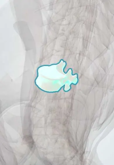
Osteoporotic vertebral compression fracture !
An osteoporotic vertebral fracture is the collapse of a vertebra weakened by osteoporosis, often after a minor strain; it causes back pain and lost height.
Updated 2026-10-29
Structures involved
Tap one to see what it is and open it in 3D.
- T12 vertebra
- L1 vertebra
- Endplates
- T11 vertebra
- L2 vertebra
- Spinal cord
- Anterior longitudinal ligament
- Posterior longitudinal ligament
In histology
In physiology
Why it happens
What it is
Collapse of the front of a vertebral body in bone weakened by osteoporosis, after little or no trauma. It is the most common osteoporotic fracture and occurs mainly at the thoracolumbar junction.
Who gets it
Very common in older adults, especially postmenopausal women. Many are not diagnosed because they cause few symptoms. As populations in Colombia and Latin America age, the burden of osteoporotic fractures is growing.
Causes
- Osteoporosis: postmenopausal, age-related or secondary
- Minor trauma: a fall from standing, lifting, bending, coughing, or no clear event
- Pathological fracture through a tumour or metastasis (an important alternative cause)
Risk factors
- Female sex, postmenopause and older age
- Previous fragility fracture
- Long-term corticosteroid use
- Low body weight, smoking and alcohol excess
- Low calcium and vitamin D, physical inactivity
- Conditions such as rheumatoid arthritis, hyperthyroidism or hypogonadism
Pathophysiology
Osteoporosis thins trabecular bone, which makes up most of the vertebral body. Axial and flexion loads crush the anterior column, giving a wedge, biconcave or crush shape; the posterior wall usually stays intact, so neurological injury is rare. Several fractures increase thoracic kyphosis and shorten the trunk.
Classification
- By shape: wedge (most common), biconcave or crush
- By severity (Genant semiquantitative method): mild, moderate or severe loss of height
- Stable (posterior wall intact) or with retropulsion of fragments into the canal
Symptoms and signs
- Sudden mid or low back pain after minor strain, worse on standing and moving
- Local tenderness over the spinous process
- Loss of height and progressive thoracic kyphosis (dowager's hump)
- Many fractures are painless and found by chance on X-ray
Diagnosis
- Clinical
- Back pain in an older person with osteoporosis risk factors, with tenderness at the fracture level. Full neurological examination and a search for red flags.
- Tests
- Calcium, phosphate, renal and liver function, vitamin D, and thyroid function. Serum protein electrophoresis to rule out myeloma, and other tests for secondary causes. Bone densitometry (DXA) once the fracture is diagnosed.
- Imaging
- Lateral X-ray shows loss of vertebral height. MRI separates recent from old fractures (oedema on STIR) and helps rule out tumour; CT shows the posterior wall and canal. DXA is part of the osteoporosis work-up.
Differential diagnosis
- Pathological fracture from metastasis or multiple myeloma
- Vertebral osteomyelitis or spinal tuberculosis
- Traumatic burst fracture
- Mechanical low back pain
- Referred visceral pain
Goal of treatment
Control pain, restore mobility quickly, prevent new fractures by treating osteoporosis, and recognise fractures caused by tumour or infection.
Treatment
- Conservative
- Short rest followed by early mobilisation; a brace is sometimes used for comfort, though evidence is limited.
- Medical
- Analgesics, chosen with care in older adults. Osteoporosis treatment with calcium, vitamin D and antiresorptive or anabolic agents (bisphosphonates and others).
- Procedure
- Vertebroplasty or kyphoplasty (cement injection into the vertebral body) for persistent pain in selected patients; evidence for benefit is mixed.
- Surgical
- Decompression and stabilisation, rarely needed: for neurological deficit, instability or severe deformity.
- Rehabilitation
- Physical therapy for posture, back extensor strength and balance, and fall prevention.
Complications
- Chronic back pain
- Progressive kyphosis and loss of height
- Reduced lung capacity and early satiety from trunk shortening
- New vertebral and hip fractures
- Loss of independence and higher mortality
Prognosis
Acute pain usually settles over weeks to a few months. A first vertebral fracture greatly increases the risk of further fractures, so treating osteoporosis is central.
Prevention
- Screening and treatment of osteoporosis
- Adequate calcium, vitamin D and protein
- Weight-bearing and resistance exercise
- Fall prevention, stopping smoking and limiting alcohol
Test yourself
Think of the answer, then tap the question to see it.
Which part of the vertebra collapses in a typical osteoporotic compression fracture?
The anterior part of the vertebral body (anterior column), giving a wedge; the posterior wall usually stays intact.
Where do osteoporotic vertebral fractures most often occur?
In the mid-thoracic spine and at the thoracolumbar junction (around T12–L1).
Which findings suggest that a vertebral fracture may be due to malignancy?
Cancer history, weight loss, night pain, fracture above T5, posterior wall involvement or destruction of the pedicles.
What postural change is caused by multiple vertebral compression fractures?
Progressive thoracic kyphosis (dowager's hump) with loss of height.
After an osteoporotic vertebral fracture, what is key to preventing new fractures?
Bone density assessment and treatment of osteoporosis, plus fall prevention.
Related conditions
- Mechanical low back pain
- Spinal cord injury
- Femoral Neck Fracture
- Intertrochanteric hip fracture
- Distal radius fracture (Colles)
- Vertebral osteomyelitis
- Ankylosing spondylitis
Educational information, not medical advice. For any symptom, see a health professional.
Sources: Vertebral Compression Fractures (StatPearls (NCBI Bookshelf), 2025).
Spotted a mistake?We are always improving for the community. Tell us what is wrong and our team will review it.Report a mistake
Representative model, for educational purposes only.