Eisenmenger syndrome !
A long-standing hole in the heart damages the lung arteries, the shunt reverses and blue blood reaches the body, causing cyanosis.
Updated 2026-11-01
Structures involved
Tap one to see what it is and open it in 3D.
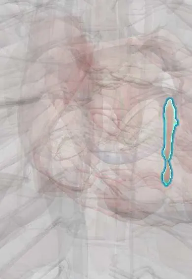
- Interventricular septum
- Pulmonary arteries
- Right ventricle
- Pulmonary trunk
- Interatrial septum
- Ligamentum arteriosum
- Left ventricle
- Right atrium
- Segmental lung vessels
Why it happens
- Hypertrophy: physiologic and pathologic
- Physiology of congenital shunts
- The pulmonary circulation and hypoxic vasoconstriction
- Polycythemia: physiological causes
- Blood vessels: artery, vein and capillary layers
What it is
Eisenmenger syndrome is the end stage of an unrepaired large left-to-right shunt (most often a ventricular septal defect, also patent ductus arteriosus, atrioventricular septal defect or a large atrial septal defect). Years of high pulmonary blood flow and pressure cause irreversible pulmonary vascular disease; when pulmonary resistance equals or exceeds systemic resistance, the shunt becomes bidirectional or right-to-left, producing cyanosis and secondary erythrocytosis.
Who gets it
It has become less common where congenital defects are repaired in infancy, but it is still seen in adults in settings with late diagnosis or limited access to paediatric cardiac surgery, including parts of Latin America. Children with Down syndrome and atrioventricular septal defects develop pulmonary vascular disease earlier. Women are particularly affected because of the very high risk of pregnancy.
Causes
- Unrepaired large ventricular septal defect (most common)
- Patent ductus arteriosus (cyanosis and clubbing of the toes but not the hands: differential cyanosis)
- Atrioventricular septal defect, especially in Down syndrome
- Large atrial septal defect or sinus venosus defect (develops later, usually in adulthood)
- Complex lesions such as truncus arteriosus or transposition with a large VSD
Risk factors
- Late diagnosis or delayed surgery of a large shunt
- Post-tricuspid shunts (VSD, PDA), which transmit systemic pressure to the lungs
- Down syndrome
- Residence at high altitude (added hypoxic pulmonary vasoconstriction)
Pathophysiology
High flow and pressure injure the pulmonary arterioles: medial hypertrophy is followed by intimal proliferation and fibrosis, then necrotising arteritis and plexiform lesions. Pulmonary vascular resistance rises progressively and becomes fixed. The right ventricle hypertrophies, and once its pressure reaches systemic levels, deoxygenated blood crosses to the systemic circulation. Chronic hypoxaemia stimulates erythropoietin, causing secondary erythrocytosis with hyperviscosity, iron deficiency, high urate and pigment gallstones, plus a bleeding and clotting tendency. Paradoxical emboli can reach the brain because venous blood bypasses the lung filter.
Classification
- By level of shunt: pre-tricuspid (atrial) vs post-tricuspid (ventricular, ductal)
- Part of pulmonary arterial hypertension associated with congenital heart disease (group 1 of the pulmonary hypertension classification)
Symptoms and signs
- Central cyanosis and digital clubbing
- Exertional dyspnoea and fatigue
- Syncope, chest pain and palpitations
- Hyperviscosity symptoms: headache, dizziness, visual disturbance, paraesthesia
- Haemoptysis
Diagnosis
- Clinical
- Cyanosis and clubbing in a patient with a known or suspected shunt; loud single P2, right ventricular heave, murmur of pulmonary or tricuspid regurgitation; the original shunt murmur often softens or disappears as pressures equalise. In PDA, cyanosis and clubbing affect the feet more than the hands.
- Tests
- Pulse oximetry at rest and on exertion; full blood count (erythrocytosis), ferritin and iron studies (iron deficiency is common), urate, renal function, coagulation. ECG: right ventricular hypertrophy and right atrial enlargement.
- Imaging
- Echocardiography identifies the defect, the shunt direction and right ventricular pressure. Right heart catheterisation measures pulmonary vascular resistance and confirms that the shunt is not repairable. Chest X-ray shows enlarged central pulmonary arteries with peripheral pruning; CT may show thrombus or calcification in dilated pulmonary arteries.
Differential diagnosis
- Idiopathic pulmonary arterial hypertension
- Chronic thromboembolic pulmonary hypertension
- Pulmonary hypertension from lung disease (COPD, interstitial lung disease)
- Uncorrected cyanotic congenital heart disease without pulmonary vascular disease
- Polycythaemia vera and other causes of erythrocytosis
Goal of treatment
Improve exercise capacity and quality of life, prevent complications of hypoxaemia and erythrocytosis, and avoid situations that destabilise the circulation; defect closure is contraindicated once the syndrome is established.
Treatment
- Medical
- Pulmonary arterial hypertension therapy: endothelin receptor antagonists (for example bosentan), phosphodiesterase-5 inhibitors and prostanoids, alone or combined.
- Medical
- Supportive care: correct iron deficiency, keep well hydrated, treat arrhythmias and heart failure; therapeutic phlebotomy only for symptomatic hyperviscosity with fluid replacement, never routinely.
- Lifestyle
- Pregnancy is contraindicated, so effective contraception counselling is essential (oestrogen-containing methods increase thrombotic risk); avoid dehydration, strenuous isometric exercise and high altitude; vaccination against influenza and pneumococcus.
- Surgical
- Lung transplantation with repair of the defect, or combined heart-lung transplantation, for selected patients with end-stage disease.
- Follow-up
- Nursing: air filters on every intravenous line to prevent paradoxical air embolism, careful fluid balance, oximetry, avoid sudden vasodilation, procedures and anaesthesia only in specialised congenital heart centres.
Complications
- Right ventricular failure
- Arrhythmias and sudden cardiac death
- Haemoptysis and pulmonary artery thrombosis
- Paradoxical embolism, ischaemic stroke and brain abscess
- Iron deficiency, hyperuricaemia with gout, pigment gallstones, renal dysfunction
- Infective endocarditis
Prognosis
Many patients survive into the third to fifth decade, longer than in idiopathic pulmonary arterial hypertension, but life expectancy is reduced and quality of life is limited. Pregnancy, non-cardiac surgery and dehydration carry high risk.
Prevention
- Early detection of murmurs and congenital heart defects (newborn and paediatric screening)
- Timely closure of large left-to-right shunts in infancy, before pulmonary resistance becomes fixed
- Early cardiology referral for children with Down syndrome
Test yourself
Think of the answer, then tap the question to see it.
Which congenital defect most commonly leads to Eisenmenger syndrome?
An unrepaired large ventricular septal defect.
What is the hallmark haemodynamic change?
Pulmonary vascular resistance reaches or exceeds systemic resistance, reversing the shunt to right-to-left.
Why do patients with a PDA and Eisenmenger syndrome have cyanosis of the toes but not the fingers?
Deoxygenated blood enters the aorta distal to the left subclavian artery (differential cyanosis).
Should the septal defect be closed once Eisenmenger syndrome is established?
No; closure is contraindicated because the defect acts as a relief valve for the right ventricle.
What nursing safety measure applies to intravenous lines in these patients?
Use air filters and remove all bubbles to prevent paradoxical air embolism.
Related conditions
- Ventricular septal defect
- Atrial septal defect
- Patent ductus arteriosus
- Truncus arteriosus, TAPVR and tricuspid atresia
- Pulmonary hypertension and cor pulmonale
- Down syndrome (trisomy 21)
- Heart failure
- Infective endocarditis
- Brain abscess
- Ischaemic stroke
- Polycythaemia vera
- Gout
Educational information, not medical advice. For any symptom, see a health professional.
Sources: Eisenmenger syndrome (MedlinePlus, US National Library of Medicine, 2026); Congenital heart disease (MedlinePlus, US National Library of Medicine, 2026).
Spotted a mistake?We are always improving for the community. Tell us what is wrong and our team will review it.Report a mistake
Representative model, for educational purposes only.