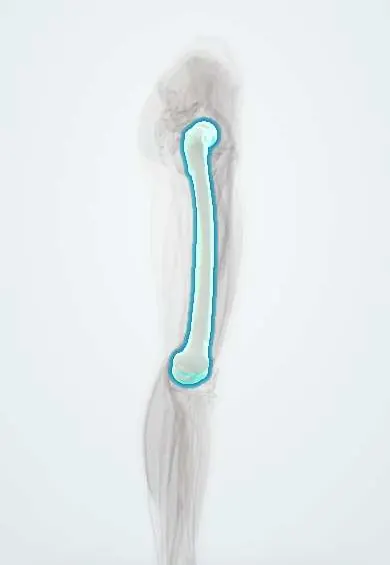
Intertrochanteric hip fracture !
An intertrochanteric fracture is a hip fracture through the top of the femur, outside the joint, typical of older adults with weak bones.
Updated 2026-10-29
Structures involved
Tap one to see what it is and open it in 3D.
Why it happens
What it is
A hip fracture through the femur between the greater and lesser trochanters, outside the joint capsule, typical of older adults with osteoporosis.
Who gets it
One of the two main types of hip fracture in older adults, mostly after a simple fall. Its frequency is rising in Colombia and Latin America as the population ages.
Causes
- Low-energy fall from standing height in older adults
- High-energy trauma in younger people
- Pathological fracture through a tumour or metastasis
Risk factors
- Osteoporosis
- Older age and female sex
- Falls: poor balance, weak muscles, poor vision, sedatives
- Dementia and frailty
- Previous fragility fracture
Pathophysiology
The region is well supplied by blood and made of cancellous bone, so avascular necrosis is rare and union is the rule if fixed well. Stability depends on the posteromedial cortex near the lesser trochanter. The iliopsoas and gluteal muscles pull the fragments, causing shortening and external rotation.
Classification
- Stable or unstable (posteromedial comminution, reverse obliquity, subtrochanteric extension)
- AO/OTA classification (31A)
- Evans classification
Symptoms and signs
- Hip or groin pain after a fall
- Inability to stand or walk
- Leg shortened and externally rotated
- Bruising over the lateral hip (later)
Diagnosis
- Clinical
- History of a fall, short externally rotated leg, pain on moving the hip. Assess the cause of the fall, cognition, skin and other injuries.
- Tests
- Blood count, electrolytes, kidney function, clotting tests, group and screen, ECG; evaluation of bone health (e.g. vitamin D, calcium) later.
- Imaging
- AP pelvis and lateral hip X-rays. MRI or CT if pain persists and X-rays are normal (occult fracture).
Differential diagnosis
- Femoral neck fracture
- Subtrochanteric fracture
- Pubic ramus or acetabular fracture
- Greater trochanter fracture
- Hip contusion
Goal of treatment
Fix the fracture early so the patient can stand and walk with full weight bearing, reduce the complications of bed rest, and prevent the next fracture.
Treatment
- Acute
- Early assessment and optimisation, analgesia (including regional nerve blocks), fluid balance and thromboprophylaxis.
- Surgical
- Surgery is preferred, ideally within about the first 48 hours: a sliding hip screw for stable patterns, an intramedullary nail for unstable ones.
- Rehabilitation
- Mobilisation from the day after surgery and orthogeriatric rehabilitation to regain walking.
- Medical
- Osteoporosis treatment (e.g. bisphosphonates) and fall prevention after the fracture.
Complications
- Pneumonia, pressure ulcers, urinary infection from immobility
- Deep vein thrombosis and pulmonary embolism
- Delirium
- Fixation failure (screw cut-out) and malunion
- Loss of independence and increased mortality
Prognosis
The fracture usually heals, but many patients do not regain their former mobility, and mortality in the first year is substantial. Early surgery and rehabilitation improve outcomes.
Prevention
- Diagnosis and treatment of osteoporosis
- Fall prevention: exercise, home safety, medication review, vision care
Test yourself
Think of the answer, then tap the question to see it.
Why is avascular necrosis rare after intertrochanteric fracture compared with femoral neck fracture?
The fracture is extracapsular through well-perfused cancellous bone, so the femoral head's blood supply is usually spared.
What is the typical position of the leg after a hip fracture?
Shortened and externally rotated.
Which implant is usually preferred for an unstable intertrochanteric fracture?
An intramedullary (cephalomedullary) nail; stable patterns are often treated with a sliding hip screw.
Why do guidelines favour surgery within about 48 hours?
Early fixation allows early mobilisation and is linked with fewer complications such as pneumonia, pressure ulcers and thrombosis.
Related conditions
- Femoral Neck Fracture
- Deep vein thrombosis
- Greater Trochanteric Pain Syndrome
- Osteoporotic vertebral compression fracture
Educational information, not medical advice. For any symptom, see a health professional.
Sources: Intertrochanteric Femur Fracture (StatPearls (NCBI Bookshelf), 2023).
Spotted a mistake?We are always improving for the community. Tell us what is wrong and our team will review it.Report a mistake
Representative model, for educational purposes only.