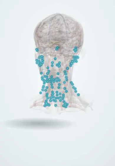
Cervical lymphadenopathy !
Cervical lymphadenopathy means swollen lymph nodes in the neck; most are a reaction to infection, but hard or growing nodes need to be checked.
Updated 2026-10-30
Structures involved
Tap one to see what it is and open it in 3D.
- Neck lymph nodes
- Sternocleidomastoid
- Internal jugular vein
- Palatine tonsils
- Thyroid gland
- Accessory nerve (XI)
In immunology
In embryology
In the clinical exam
Why it happens
- Granulomatous inflammation: caseating vs non-caseating, the causes to know
- Pathways of spread and the usual metastatic sites
- Biopsy: excisional, incisional, core needle, punch and fine-needle aspiration
- Cytology: the Pap test, fine-needle aspiration and body fluids
- Lymph node and spleen under the microscope
What it is
Enlargement of the lymph nodes of the neck. Most cases are reactive to infection and resolve; persistent, hard or growing nodes need evaluation for tuberculosis, lymphoma or metastatic cancer.
Who gets it
Very common, especially in children with respiratory infections. Tuberculosis is a key cause in Latin America: in adults biopsied for unexplained lymphadenopathy in three public hospitals in Lima, Peru, 34% had tuberculosis, 13% lymphoma and 14% other cancers. In older adults and smokers, metastatic cancer is a key concern.
Causes
- Reactive: viral and bacterial upper respiratory, dental and skin infections
- Infectious: EBV and CMV, tuberculosis, HIV, toxoplasmosis, cat-scratch disease
- Malignant: lymphoma and metastases (head and neck squamous cell, thyroid, skin)
- Inflammatory: Kawasaki disease, sarcoidosis, autoimmune diseases
Risk factors
- Recent infection of throat, ear, teeth or scalp
- Tuberculosis exposure and HIV infection
- Tobacco and alcohol use (head and neck cancer)
- Older age
Pathophysiology
Nodes enlarge when lymphocytes and macrophages multiply in response to antigens draining from their territory, or when inflammatory, infected or malignant cells infiltrate them. The location of the node points to its drainage area: for example the supraclavicular node on the left reflects abdominal and thoracic disease.
Classification
- Localised versus generalised
- Acute versus persistent (longer than a few weeks)
- By neck level (I–VI) and supraclavicular nodes
Symptoms and signs
- Palpable lump in the neck
- Tender, soft, mobile nodes in reactive or infective cases
- Hard, fixed, painless nodes in malignancy
- Matted nodes or a draining sinus in tuberculosis (scrofula)
- Fever, night sweats and weight loss in systemic disease
Diagnosis
- Clinical
- History and full head and neck examination, including mouth, pharynx, skin and other node groups, liver and spleen.
- Tests
- Blood count and film, inflammatory markers, serology (EBV, HIV, toxoplasma), tuberculin or interferon-gamma test; fine-needle aspiration or core or excisional biopsy of persistent nodes.
- Imaging
- Ultrasound of the neck; CT or PET for suspected malignancy; chest radiograph.
Differential diagnosis
- Thyroid nodule
- Branchial cleft or thyroglossal duct cyst
- Salivary gland swelling
- Lipoma
- Carotid body tumour
Goal of treatment
Find and treat the underlying cause; avoid missing malignancy or tuberculosis.
Treatment
- Follow-up
- Observation and review in a few weeks for small, soft, likely reactive nodes.
- Medical
- Treatment of the cause: antibiotics for bacterial lymphadenitis, antituberculosis therapy, or antiretroviral therapy.
- Surgical
- Excisional biopsy when lymphoma is suspected; drainage of a suppurative node; neck dissection for metastatic cancer.
Complications
- Abscess formation
- Sinus tract (tuberculosis)
- Delayed diagnosis of cancer
Prognosis
Reactive nodes usually shrink within weeks. Prognosis otherwise depends on the cause.
Test yourself
Think of the answer, then tap the question to see it.
Which neck node finding is most worrying for malignancy?
A hard, fixed, painless or enlarging node, especially in the supraclavicular fossa.
What is Virchow node?
An enlarged left supraclavicular node, classically from abdominal malignancy such as gastric cancer.
What is scrofula?
Tuberculous cervical lymphadenitis, often with matted nodes and draining sinuses.
Which biopsy is preferred when lymphoma is suspected?
Excisional (or core) biopsy, which preserves node architecture; fine-needle aspiration alone is often not enough.
Related conditions
- Streptococcal pharyngitis
- Tuberculosis
- HIV infection and AIDS
- Thyroid cancer (papillary)
- Deep neck infection and Ludwig angina
- Mumps
- Laryngeal cancer
- Thyroid nodule
Educational information, not medical advice. For any symptom, see a health professional.
Sources: Lymphadenopathy (StatPearls (NCBI Bookshelf), 2026); Histological Examination in Obtaining a Diagnosis in Patients with Lymphadenopathy in Lima, Peru (The American journal of tropical medicine and hygiene, 2017) · adapted, CC BY 4.0.
Spotted a mistake?We are always improving for the community. Tell us what is wrong and our team will review it.Report a mistake
Representative model, for educational purposes only.