Osteopetrosis !
Inherited "marble bone" disease: bones become very dense but brittle because they are not remodelled, which can crush nerves and crowd out the marrow.
Updated 2026-11-01
Structures involved
Tap one to see what it is and open it in 3D.
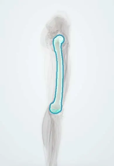
- Femur
- Temporal bones
- Optic canal
- Optic nerve
- Facial nerve (VII)
- Vestibulocochlear nerve (VIII)
- Cochlea
- Mandible
- Tibia
- Hip bone
Why it happens
- Mendelian inheritance: autosomal dominant, recessive and X-linked
- Bone remodeling
- Bone tissue: cells, compact and spongy bone
- Hematopoiesis and bone marrow
What it is
A group of inherited disorders in which osteoclasts fail to resorb bone. Bone keeps being formed but is not remodelled, so the skeleton becomes dense yet brittle, marrow space shrinks and the skull foramina narrow. Severity ranges from a lethal infantile form to an adult form found by chance on X-ray.
Who gets it
The autosomal dominant adult form affects about 1 in 20,000 people and is the most common. The severe autosomal recessive infantile form affects roughly 1 in 250,000, and is more frequent where consanguineous marriage is common.
Causes
- Autosomal dominant osteopetrosis (Albers-Schönberg disease): mostly CLCN7 variants
- Autosomal recessive (infantile, "malignant") osteopetrosis: TCIRG1 variants in about half, also CLCN7, OSTM1, TNFSF11 (RANKL) and TNFRSF11A (RANK)
- Carbonic anhydrase II deficiency: osteopetrosis with renal tubular acidosis and cerebral calcification
- Intermediate forms with either inheritance pattern
Risk factors
- Family history of osteopetrosis
- Parental consanguinity (recessive forms)
Pathophysiology
Osteoclasts need acidification and chloride transport at the bone surface to dissolve mineral; when this fails, primary spongiosa and calcified cartilage persist. Bone becomes dense but poorly organised, so it fractures easily and heals slowly. Narrowed cranial foramina compress the optic, facial and auditory nerves. Loss of marrow space causes pancytopenia with extramedullary haematopoiesis in the liver and spleen. Calcium cannot be mobilised from bone, so infants may develop hypocalcaemia.
Classification
- Autosomal recessive (severe, infantile)
- Intermediate autosomal osteopetrosis
- Autosomal dominant (adult, benign, Albers-Schönberg)
- Carbonic anhydrase II deficiency
Symptoms and signs
- Fractures after minor trauma; bone pain and hip osteoarthritis in adults
- Vision loss, deafness and facial palsy from cranial nerve compression
- Anaemia, bleeding and repeated infections from marrow failure (infantile form)
- Enlarged liver and spleen, failure to thrive, short stature, macrocephaly
- Dental problems and osteomyelitis of the jaw
Diagnosis
- Clinical
- Fractures, cranial nerve palsies and cytopenias in a child, or an incidental finding of dense bones in an adult; family history.
- Tests
- Full blood count and blood film (anaemia, thrombocytopenia, leucoerythroblastic picture); calcium, phosphate and PTH; acid phosphatase and creatine kinase BB may be raised. Genetic testing confirms the type. Eye examination and hearing tests.
- Imaging
- Radiographs show diffusely dense bones, "bone-in-bone" appearance, "sandwich" (rugger-jersey) vertebrae, club-shaped metaphyses and a thick skull base; CT or MRI of the skull assesses foramina and hydrocephalus.
Differential diagnosis
- Pyknodysostosis
- Paget disease of bone
- Osteosclerotic bone metastases (prostate, breast)
- Fluorosis and other toxic osteosclerosis
- Myelofibrosis with osteosclerosis
Goal of treatment
In severe infantile forms, restore osteoclast function early with haematopoietic stem cell transplantation before vision is lost; in all forms, treat fractures, nerve compression, marrow failure and infections.
Treatment
- Procedure
- Haematopoietic stem cell transplantation for severe osteoclast-intrinsic recessive forms, because osteoclasts come from marrow precursors; it is not useful in RANKL-deficient forms.
- Medical
- Calcium and vitamin D management for hypocalcaemia; transfusions and treatment of infections; interferon gamma-1b has been used in selected patients.
- Surgical
- Fracture fixation (technically difficult because of hard, brittle bone); decompression of the optic canal in selected cases; joint replacement for hip arthritis.
- Conservative
- Careful dental care and oral hygiene to prevent jaw osteomyelitis.
- Follow-up
- Regular blood counts, vision and hearing checks, and growth monitoring in children.
Complications
- Recurrent fractures and delayed healing
- Blindness, deafness and facial nerve palsy
- Pancytopenia with infections and bleeding
- Osteomyelitis, especially of the mandible
- Hypocalcaemia and hydrocephalus in infants
Prognosis
Untreated severe infantile osteopetrosis is usually fatal in early childhood; transplantation can cure the bone and marrow disease if done early, but vision already lost does not return. The adult dominant form usually allows a normal lifespan, with morbidity from fractures, arthritis and jaw infection.
Test yourself
Think of the answer, then tap the question to see it.
What cellular defect causes osteopetrosis?
Failure of osteoclast bone resorption (for example, defective acidification of the resorption pit).
Why does severe infantile osteopetrosis cause pancytopenia and hepatosplenomegaly?
Dense bone obliterates marrow space, so blood formation moves to the liver and spleen.
Why can stem cell transplantation cure the severe recessive form?
Osteoclasts derive from haematopoietic precursors, so donor marrow supplies working osteoclasts.
Which cranial nerves are typically compressed in osteopetrosis?
The optic (II), facial (VII) and vestibulocochlear (VIII) nerves, through narrowed foramina.
Which enzyme deficiency combines osteopetrosis with renal tubular acidosis and cerebral calcification?
Carbonic anhydrase II deficiency.
Related conditions
- Osteomyelitis
- Aplastic anaemia
- Hearing loss (conductive and sensorineural, including sudden hearing loss)
- Bell palsy
- Hypocalcaemia
- Renal tubular acidosis
- Osteogenesis imperfecta
- Paget disease of bone
- Essential thrombocythaemia and primary myelofibrosis
- Femoral Neck Fracture
Educational information, not medical advice. For any symptom, see a health professional.
Sources: Osteopetrosis: MedlinePlus Genetics (MedlinePlus Genetics (U.S. National Library of Medicine), 2025).
Spotted a mistake?We are always improving for the community. Tell us what is wrong and our team will review it.Report a mistake
Representative model, for educational purposes only.