Conditions · Metabolism (biochemistry)
Respiratory acidosis !
The lungs do not remove enough CO₂ and the blood turns acidic; it happens in COPD, sedative overdose and muscle weakness.
Updated 2026-11-01
Structures involved
Tap one to see what it is and open it in 3D.
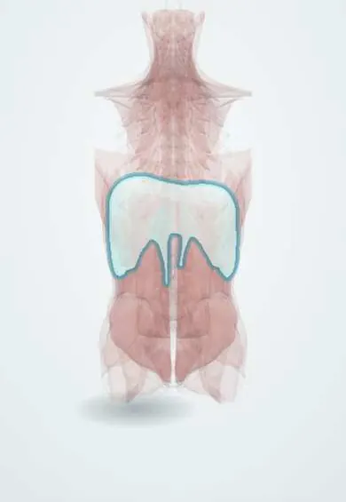
- Diaphragm
- Medulla oblongata
- Right upper lobe
- Right middle lobe
- Right lower lobe
- Left upper lobe
- Left lower lobe
- Main bronchi
- Phrenic nerves
In biochemistry
In the clinical exam
In physiology
- Reading an arterial blood gas step by step
- Respiratory acidosis and alkalosis
- Buffers and the Henderson–Hasselbalch equation
- Mixed acid–base disorders
- Dead space and alveolar ventilation
- CO₂ transport and the Haldane effect
- Control of breathing, chemoreceptors and lung reflexes
- Oxygenation: SpO₂, PaO₂, cyanosis and oxygen therapy
- Hypoxemic and hypercapnic respiratory failure
What it is
A primary rise in pCO₂ (hypercapnia) from alveolar hypoventilation, with a fall in pH; it may be acute or chronic and kidney-compensated.
Who gets it
Common in COPD exacerbations, a highly prevalent disease in Colombia from smoking and wood-smoke exposure, and in opioid or sedative poisoning. Severe obesity adds cases of chronic hypoventilation.
Causes
- Depressed respiratory centre: opioids, benzodiazepines, alcohol, anaesthesia, brainstem injury
- Neuromuscular disease: Guillain-Barré, myasthenia, high spinal cord injury, amyotrophic lateral sclerosis
- Lung or airway disease: COPD, severe asthma, extensive pneumonia, advanced pulmonary oedema
- Chest wall: obesity (obesity hypoventilation syndrome), kyphoscoliosis, flail chest
- Inadequate mechanical ventilation
Risk factors
- COPD from tobacco or wood smoke
- Opioid or sedative use
- Severe obesity and sleep apnoea
- Neuromuscular diseases
Pathophysiology
CO₂ production outpaces its removal and pCO₂ rises; through the carbonic anhydrase reaction H⁺ and HCO₃⁻ increase. In the acute form only cell buffers act and HCO₃⁻ rises just 1 mmol/L per 10 mmHg; in the chronic form the kidney increases H⁺ and ammonium excretion and reabsorbs more HCO₃⁻ (about 3.5–4 per 10 mmHg) over 3–5 days. CO₂ crosses the blood–brain barrier, acidifies cerebrospinal fluid and dilates cerebral vessels: headache, drowsiness and, in severe cases, coma from CO₂ narcosis. On room air it is nearly always accompanied by hypoxaemia.
Classification
- Acute: low pH with near-normal HCO₃⁻
- Chronic: near-normal pH with high HCO₃⁻
- Acute-on-chronic: COPD exacerbation
Symptoms and signs
- Breathlessness or, conversely, slow shallow breathing (sedatives)
- Morning headache (chronic hypoventilation)
- Drowsiness, confusion
- Flapping tremor (asterixis)
- Warm, flushed skin from vasodilatation
Diagnosis
- Clinical
- History of COPD, sedatives or muscle weakness; respiratory rate, accessory muscle use and level of consciousness.
- Tests
- Arterial gases (high pCO₂, low pH, HCO₃⁻ by chronicity), oximetry, electrolytes, toxicology, lung or neuromuscular function tests once stable.
- Imaging
- Chest X-ray for pneumonia, oedema or pneumothorax; CT according to the cause.
Treatment
- Acute
- Secure the airway and ventilation; opioid antagonist (naloxone) for overdose
- Procedure
- Non-invasive ventilation in COPD exacerbations or obesity hypoventilation; intubation and mechanical ventilation if it fails
- Medical
- Treat the cause: bronchodilators and corticosteroids in COPD or asthma, antibiotics in pneumonia, immunotherapy in Guillain-Barré
- Follow-up
- Controlled oxygen with a moderate saturation target in COPD to avoid worsening hypercapnia; slow correction of chronic pCO₂
Complications
- Coma from CO₂ narcosis
- Arrhythmias
- Respiratory arrest
- Post-hypercapnic alkalosis if over-ventilated
Prognosis
Depends on the cause; acute hypercapnia needing ventilation in COPD marks advanced disease.
Prevention
- Stopping smoking and reducing wood-smoke exposure
- Careful use of opioids and sedatives
- Treating sleep apnoea
Test yourself
Think of the answer, then tap the question to see it.
What separates acute from chronic respiratory acidosis?
HCO₃⁻: it rises 1 mmol/L per 10 mmHg of pCO₂ in the acute form and 3.5–4 in the chronic form, thanks to renal compensation.
Why can uncontrolled oxygen worsen hypercapnia in COPD?
It worsens ventilation–perfusion mismatch and the Haldane effect, and in some patients it reduces respiratory drive.
What causes headache and drowsiness in hypercapnia?
CO₂ crosses the blood–brain barrier, acidifies cerebrospinal fluid and dilates cerebral vessels.
Which drugs cause acute respiratory acidosis by central depression?
Opioids, benzodiazepines and other sedatives, as well as alcohol.
Related conditions
- Respiratory alkalosis
- Chronic obstructive pulmonary disease (COPD)
- Obstructive sleep apnoea
- Guillain-Barré syndrome
- Asthma
- Obesity
Educational information, not medical advice. For any symptom, see a health professional.
Sources: Respiratory acidosis: MedlinePlus Medical Encyclopedia (MedlinePlus, US National Library of Medicine, 2026); Arterial Blood Gas (ABG) Test: MedlinePlus Medical Test (MedlinePlus, US National Library of Medicine, 2026); Textbook of Biochemistry for Medical Students, 6th ed. (Jaypee Brothers Medical Publishers (Vasudevan DM, Sreekumari S, Vaidyanathan K), 2011).
Spotted a mistake?We are always improving for the community. Tell us what is wrong and our team will review it.Report a mistake
Representative model, for educational purposes only.