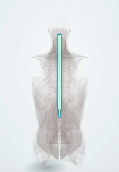
Spinal muscular atrophy !
Spinal muscular atrophy is an inherited disease in which the nerve cells that move muscles die, causing weakness. New treatments work best when started early.
Updated 2026-11-01
Structures involved
Tap one to see what it is and open it in 3D.
Why it happens
- Mendelian inheritance: autosomal dominant, recessive and X-linked
- Atrophy
- Upper versus lower motor neuron lesions
- RNA processing (cap, poly-A tail and splicing)
What it is
An autosomal recessive motor neuron disease in which the anterior horn cells of the spinal cord and motor nuclei of the brainstem degenerate, causing symmetric, proximal-predominant weakness, hypotonia and muscle wasting. It was the most common genetic cause of infant death before disease-modifying treatment.
Who gets it
About 1 in 8,000 to 10,000 births worldwide; about 1 in 40 to 60 people carry an SMN1 deletion. Type 1 is the most common form. In Colombia and Latin America, access to newborn screening and to high-cost disease-modifying drugs is still uneven, so many children are diagnosed only after symptoms appear.
Causes
- Homozygous deletion or mutation of the SMN1 gene (chromosome 5q13) in about 95% of cases
- The number of copies of the neighbouring SMN2 gene modifies severity: more copies, milder disease
- Autosomal recessive inheritance; both parents are usually healthy carriers
Risk factors
- Both parents being carriers (25% risk in each pregnancy)
- Family history of SMA or of unexplained infant deaths with weakness
- Consanguinity
Pathophysiology
The SMN (survival motor neuron) protein is needed for motor neuron survival. SMN1 makes most of the working protein; SMN2 differs by one base that causes most of its transcripts to skip exon 7, producing a small amount of functional protein. Without SMN1, anterior horn cells die, muscles lose their nerve supply and waste, giving lower motor neuron signs with intact sensation and cognition.
Classification
- Type 0: onset before birth, severe weakness and respiratory failure at birth
- Type 1 (Werdnig-Hoffmann): onset before 6 months, never sits unaided
- Type 2: onset 6–18 months, sits but never walks independently
- Type 3 (Kugelberg-Welander): onset after 18 months, walks but may lose the ability later
- Type 4: adult onset, mild weakness
Symptoms and signs
- Floppy infant: marked hypotonia with frog-leg posture
- Symmetric weakness, worse in proximal muscles and legs than arms
- Absent or reduced deep tendon reflexes
- Tongue fasciculations and fine hand tremor
- Weak cry, poor feeding and swallowing difficulty
- Bell-shaped chest and paradoxical (abdominal) breathing from intercostal weakness with a spared diaphragm
- Alert face with normal cognition and sensation; delayed or lost motor milestones
Diagnosis
- Clinical
- Suspected in a hypotonic infant or child with symmetric proximal weakness, absent reflexes and tongue fasciculations but normal alertness and sensation. Nursing assessment focuses on breathing, cough, swallowing, nutrition and positioning.
- Tests
- Genetic testing for homozygous SMN1 deletion is the key test, with SMN2 copy number to predict severity. Newborn screening detects it before symptoms where available. Creatine kinase is normal or mildly raised; electromyography shows denervation if genetics is unclear.
- Imaging
- Not needed for diagnosis. Spine X-rays to follow scoliosis and hip films for subluxation; chest X-ray during respiratory infections.
Differential diagnosis
- Duchenne and other muscular dystrophies (high CK, calf pseudohypertrophy)
- Congenital myopathies and congenital myasthenic syndromes
- Infant botulism
- Central hypotonia (cerebral palsy, Prader-Willi syndrome), which keeps reflexes and has other brain signs
- Pompe disease and peripheral neuropathies
Goal of treatment
Start disease-modifying treatment as early as possible, ideally before symptoms, and protect breathing, nutrition, mobility and the spine to maximise function and survival.
Treatment
- Medical
- Disease-modifying therapies: an intrathecal antisense oligonucleotide (nusinersen) and an oral small molecule (risdiplam) that increase SMN protein from the SMN2 gene.
- Medical
- Single-dose gene replacement therapy (onasemnogene abeparvovec) that delivers a working SMN1 gene, used mainly in young children.
- Follow-up
- Respiratory care: monitoring of breathing and cough, airway clearance, non-invasive ventilation and immunisations; nutritional support with gastrostomy when swallowing is unsafe.
- Rehabilitation
- Physical therapy, stretching, orthoses and wheelchairs to keep mobility and prevent contractures.
- Surgical
- Spinal fusion for progressive scoliosis.
Complications
- Respiratory failure and recurrent pneumonia, the main cause of death
- Aspiration, malnutrition and failure to thrive
- Scoliosis, hip dislocation and joint contractures
- Osteoporosis and fractures
Prognosis
Without treatment, type 1 is usually fatal or needs permanent ventilation within the first two years, while types 3 and 4 have a near-normal life span. Disease-modifying therapies have greatly changed outcomes, and children treated before symptoms may sit, stand or walk.
Prevention
- Carrier screening and genetic counselling for families at risk
- Newborn screening to start treatment before symptoms
- Prenatal diagnosis when both parents are known carriers
Test yourself
Think of the answer, then tap the question to see it.
Which gene is deleted in most cases of spinal muscular atrophy?
SMN1 on chromosome 5q, with autosomal recessive inheritance.
What modifies the severity of SMA?
The number of SMN2 gene copies: more copies produce more SMN protein and milder disease.
Which cells degenerate in SMA?
The anterior horn cells (lower motor neurons) of the spinal cord and brainstem motor nuclei.
What is the classic presentation of SMA type 1?
A floppy infant under 6 months with proximal weakness, absent reflexes, tongue fasciculations and normal alertness.
What is the main cause of death in SMA?
Respiratory failure and pneumonia from weak respiratory and swallowing muscles.
Related conditions
- Amyotrophic lateral sclerosis
- Guillain-Barré syndrome
- Myasthenia gravis
- Cerebral palsy
- Scoliosis
- Acute respiratory failure (hypoxaemic and hypercapnic)
- Inflammatory myopathies (dermatomyositis, polymyositis)
Educational information, not medical advice. For any symptom, see a health professional.
Sources: Spinal muscular atrophy: MedlinePlus Genetics (MedlinePlus Genetics, US National Library of Medicine, 2026).
Spotted a mistake?We are always improving for the community. Tell us what is wrong and our team will review it.Report a mistake
Representative model, for educational purposes only.