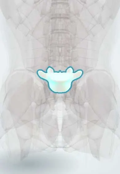
Spondylolisthesis !
Spondylolisthesis is the forward slip of one vertebra over the one below, most often in the lower back; it can cause back or leg pain.
Updated 2026-10-29
Structures involved
Tap one to see what it is and open it in 3D.
- L5 vertebra
- L4 vertebra
- Sacrum
- Low-back facet joints
- L4–L5 disc
- L5–S1 disc
- L5 nerve root
- L4 nerve root
- Cauda equina
- Ligamentum flavum
What it is
Forward slip of one vertebral body over the one below, most often L5 on S1 or L4 on L5. It may cause low back pain, radicular leg pain or neurogenic claudication, or none at all.
Who gets it
Isthmic spondylolisthesis usually starts in childhood or adolescence, often in sports with repeated spinal extension. Degenerative spondylolisthesis appears after about 50 years and is more frequent in women, mostly at L4–L5.
Causes
- Isthmic: a defect or stress fracture of the pars interarticularis (spondylolysis)
- Degenerative: facet joint arthritis and disc degeneration allow segmental instability
- Dysplastic (congenital): malformed facets or upper sacrum
- Traumatic: acute fracture of the posterior elements other than the pars
- Pathological: tumour, infection or bone disease; iatrogenic after decompression surgery
Risk factors
- Sports with repeated hyperextension: gymnastics, diving, weightlifting, football (isthmic)
- Adolescence and growth spurts (isthmic)
- Age over 50 and female sex (degenerative)
- Sagittally oriented facet joints (degenerative)
- Family history
Pathophysiology
In the isthmic type a fatigue fracture of the pars disconnects the posterior arch, and the vertebral body can drift forward. In the degenerative type the posterior arch is intact; facet arthritis and disc collapse let the whole vertebra slide, narrowing the canal and foramina. Slip and instability compress the cauda equina or the exiting and traversing roots.
Classification
- Wiltse–Newman types: dysplastic, isthmic, degenerative, traumatic, pathological, iatrogenic
- Meyerding grade by slip of the upper vertebra: grade I up to 25 %, II 26–50 %, III 51–75 %, IV 76–100 %, V (spondyloptosis) beyond 100 %
- Low grade (I–II) versus high grade (III–V)
Symptoms and signs
- Mechanical low back pain worse with extension and standing
- Radicular leg pain, numbness or weakness (often L5 in isthmic L5–S1 slips)
- Neurogenic claudication: leg pain or heaviness on walking, relieved by sitting or bending forward (degenerative)
- Hamstring tightness and altered gait or posture in children with higher-grade slips
- Palpable step-off of the spinous processes in severe slips
- Many patients have no symptoms
Diagnosis
- Clinical
- Back pain on extension, a positive single-leg extension (stork) test in adolescents with spondylolysis, and a neurological examination of the lower limbs.
- Tests
- Laboratory tests are normal and are ordered only if infection or tumour is suspected.
- Imaging
- Standing lateral X-ray shows and grades the slip; flexion–extension views show instability. Oblique views may show the pars defect as a collar on the «Scottie dog». CT defines the pars defect; MRI shows nerve compression and early stress reaction.
Differential diagnosis
- Mechanical low back pain
- Lumbar disc herniation
- Lumbar spinal stenosis without slip
- Vascular claudication from peripheral artery disease
- Vertebral infection or tumour
Goal of treatment
Control pain, keep the patient active and protect the nerves; surgery stabilises and decompresses the segment when deficit, instability, high-grade slip or persistent pain call for it.
Treatment
- Conservative
- Activity modification and temporary rest from the sport causing hyperextension; some clinicians use a brace for acute spondylolysis in adolescents.
- Medical
- Analgesics and NSAIDs; epidural steroid injection for radicular pain in selected patients.
- Rehabilitation
- Physical therapy with core stabilisation and flexion-based exercise.
- Surgical
- Surgery (decompression with or without fusion; pars repair in the young) for progressive deficit, cauda equina syndrome, high-grade or progressing slips, or refractory pain; indications vary.
Complications
- Chronic low back pain
- Radiculopathy and neurogenic claudication
- Cauda equina syndrome (rare)
- Slip progression, mainly in high-grade slips during growth
- Surgical complications: non-union, adjacent segment disease, nerve injury
Prognosis
Most low-grade slips respond to conservative care and progress little in adults. Higher-grade slips in growing children are more likely to progress and to need surgery.
Prevention
- Supervised technique and load control in sports with repeated hyperextension
- Early evaluation of persistent back pain in young athletes
- Core strengthening
Test yourself
Think of the answer, then tap the question to see it.
What is spondylolysis?
A defect or stress fracture of the pars interarticularis, usually at L5, without necessarily any slip; it is the cause of isthmic spondylolisthesis.
What does the «Scottie dog» sign show on oblique lumbar X-rays?
A spondylolysis: the pars defect appears as a collar or break across the neck of the «Scottie dog».
How does the Meyerding classification grade spondylolisthesis?
By the percentage of slip of the upper vertebra: I up to 25 %, II 26–50 %, III 51–75 %, IV 76–100 %, V beyond 100 % (spondyloptosis).
Which level and population are typical of degenerative spondylolisthesis?
L4–L5 in adults over about 50, more often women, with an intact pars and facet joint arthritis.
Which level is most typical of isthmic spondylolisthesis?
L5 on S1, often in young people who practice sports with repeated hyperextension.
Related conditions
- Lumbar spinal stenosis
- Mechanical low back pain
- Sciatica (Lumbar Radiculopathy)
- Lumbar Disc Herniation
- Cauda equina syndrome
Educational information, not medical advice. For any symptom, see a health professional.
Sources: Spondylolisthesis (StatPearls (NCBI Bookshelf), 2025).
Spotted a mistake?We are always improving for the community. Tell us what is wrong and our team will review it.Report a mistake
Representative model, for educational purposes only.