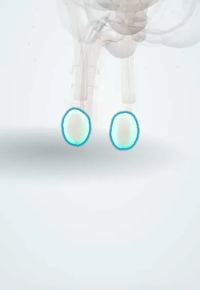
Testicular cancer !
Testicular cancer is a cancer of the testis, usually felt as a painless lump; it mostly affects young men and is highly curable even when it has spread.
Updated 2026-10-30
Structures involved
Tap one to see what it is and open it in 3D.
In biochemistry
In histology
In embryology
In the clinical exam
In physiology
Why it happens
- Chromosome abnormalities: number and structure
- Tumour markers: serum and tissue, and what each one is for
- Naming tumours: -oma, carcinoma, sarcoma, and the misleading names
What it is
Malignant tumour of the testis, most often a germ cell tumour (seminoma or non-seminoma). It usually presents as a painless lump and is highly curable even when it has spread.
Who gets it
The most common solid cancer in men roughly 15 to 35 years old, although rare overall, and incidence has risen in many countries. In Colombia, testicular cancer mortality rose 1.8% a year from 1997 to 2019 (4.3% a year at ages 15–29). Late diagnosis remains a challenge in parts of Latin America.
Causes
- Germ cell tumours (the large majority): seminoma and non-seminomas (embryonal carcinoma, yolk sac tumour, choriocarcinoma, teratoma, mixed)
- Most germ cell tumours arise from germ cell neoplasia in situ
- Sex cord–stromal tumours (Leydig cell, Sertoli cell)
- Lymphoma of the testis, mainly in older men
Risk factors
- Cryptorchidism (undescended testis), including the contralateral testis
- Previous testicular cancer
- Testicular cancer in a father or brother
- Infertility or testicular dysgenesis
- Klinefelter syndrome (mainly mediastinal germ cell tumours)
- HIV infection
Pathophysiology
Germ cell neoplasia in situ develops from fetal germ cells and progresses to invasive tumour; gain of chromosome 12p is characteristic. Spread is lymphatic along the testicular vessels to retroperitoneal (para-aortic) nodes, then haematogenous, mainly to the lungs. Some tumours secrete AFP or hCG.
Classification
- Seminoma or non-seminomatous germ cell tumour
- TNM staging with an S category for serum tumour markers
- Stages I (testis only), II (retroperitoneal nodes) and III (distant spread or very high markers)
- Prognostic risk groups for metastatic disease (good, intermediate, poor)
Symptoms and signs
- Painless, firm lump or enlargement of one testis
- Feeling of heaviness or dull ache in the scrotum or lower abdomen
- Sudden pain in some cases, from bleeding or infarction in the tumour
- Gynaecomastia or breast tenderness (hCG-secreting tumours)
- Back or flank pain from retroperitoneal lymph nodes
- Cough, dyspnoea or haemoptysis from lung metastases
Diagnosis
- Clinical
- Examination of both testes, abdomen, supraclavicular nodes and breasts. Any solid intratesticular mass is treated as cancer until proven otherwise.
- Tests
- Serum AFP, beta-hCG and LDH before and after orchiectomy; they help diagnose, stage and follow the disease. Pure seminoma does not raise AFP. Semen analysis and sperm banking are offered.
- Imaging
- Scrotal ultrasound is first-line and shows a hypoechoic intratesticular mass. CT of the chest, abdomen and pelvis stages the disease; brain imaging when indicated.
Differential diagnosis
- Epididymitis or orchitis
- Hydrocele
- Spermatocele or epididymal cyst
- Testicular torsion
- Varicocele
- Testicular haematoma after trauma
Goal of treatment
Remove the primary tumour, stage the disease and cure it with the least long-term toxicity, while preserving fertility and hormonal function where possible.
Treatment
- Surgical
- Radical inguinal orchiectomy with high ligation of the spermatic cord; a scrotal approach or biopsy is avoided. A testicular prosthesis may be offered.
- Follow-up
- Active surveillance for many patients with stage I disease, with markers and imaging at set intervals.
- Medical
- Platinum-based combination chemotherapy for metastatic or high-risk disease.
- Procedure
- Radiotherapy to the retroperitoneal nodes in selected seminomas, which are very radiosensitive.
- Surgical
- Retroperitoneal lymph node dissection in selected non-seminomas or for residual masses after chemotherapy.
Drugs for this condition
Complications
- Metastatic spread (retroperitoneum, lungs, liver, brain)
- Infertility, from the disease or its treatment
- Hypogonadism
- Late effects of chemotherapy and radiotherapy: cardiovascular disease, second cancers, hearing loss, neuropathy, kidney damage
- Retrograde ejaculation after lymph node dissection
Prognosis
One of the most curable solid cancers: survival is very high in early stages and remains high in most metastatic cases. Long-term follow-up watches for relapse and late treatment effects.
Prevention
- Early orchiopexy for undescended testes (lowers risk and makes examination easier)
- Awareness of testicular changes; routine screening of men without symptoms is not recommended by several guidelines
Test yourself
Think of the answer, then tap the question to see it.
Which serum tumour marker is never raised in pure seminoma?
Alpha-fetoprotein (AFP). A raised AFP means a non-seminomatous component.
Why is a testicular cancer removed through an inguinal and not a scrotal incision?
A scrotal approach can alter the lymphatic drainage and seed the tumour; the cord is ligated high through the inguinal canal.
To which lymph nodes does testicular cancer first spread?
Retroperitoneal (para-aortic) nodes, following the testicular vessels, not the inguinal nodes.
What is the strongest well-established risk factor for testicular cancer?
Cryptorchidism, which also raises risk in the contralateral testis.
Why can testicular cancer cause gynaecomastia?
Some tumours secrete hCG, which stimulates oestrogen production.
Related conditions
Educational information, not medical advice. For any symptom, see a health professional.
Sources: Testicular Cancer (StatPearls (NCBI Bookshelf), 2023); Testicular cancer mortality in Latin America and the Caribbean: trend analysis from 1997 to 2019 (BMC cancer, 2023) · adapted, CC BY 4.0.
Spotted a mistake?We are always improving for the community. Tell us what is wrong and our team will review it.Report a mistake
Representative model, for educational purposes only.