
Thermal tumour ablation (radiofrequency and microwave)
A needle placed in a tumour under imaging heats it until the cells die, so small cancers of the liver, kidney or lung can be treated without surgery.
What it is
Image-guided destruction of a tumour by heat: a needle electrode (radiofrequency) or antenna (microwave) is placed through the skin, or at laparoscopy or open surgery, and the tumour plus a margin of normal tissue undergo coagulative necrosis. Cryoablation (freezing) is the cold alternative.
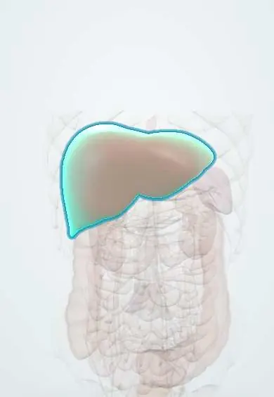
Who gets it
With resection and transplantation, ablation is one of the treatments that can cure early hepatocellular carcinoma. It is also an established option for small renal masses in patients who cannot have or decline surgery, and for medically inoperable early non-small cell lung cancer and lung metastases.
Causes
- Early hepatocellular carcinoma when resection or transplantation is not feasible or advisable, especially central tumours that would need too much liver removed
- Limited liver metastases, and recurrence after resection or a previous ablation
- Small renal cell carcinoma (T1a, up to 4 cm) in patients who cannot undergo or decline nephrectomy or general anaesthesia
- Medically inoperable early-stage non-small cell lung cancer, as an alternative to stereotactic body radiotherapy
- Lung metastases in selected patients who are unsuitable for surgery or need lung to be preserved
Risk factors
- Tumour larger than about 3 cm: local control falls as size increases
- Tumour beside a large vessel: blood flow carries heat away (heat sink) and can leave viable tumour
- Tumour next to bile ducts, diaphragm or bowel (liver), the renal hilum or proximal ureter (kidney), or the pleura (lung)
- Cirrhosis with coagulopathy (bleeding); biliary–enteric anastomosis, diabetes or oil retained after chemoembolisation (abscess)
- In the lung: emphysema, a long path through aerated lung, older age and several tumours treated (pneumothorax)
Pathophysiology
Radiofrequency current (frictional heating) or microwaves (agitation of water molecules) raise tissue temperature → proteins coagulate and cells die (coagulative necrosis) in a zone around the needle → the zone must cover the tumour plus a margin of normal tissue → blood flow in nearby large vessels cools the edge (heat sink), more so with radiofrequency, so the margin may survive → structures inside the zone are injured as well (bile ducts, bowel, ureter, pleura) → the dead tissue is not removed, so success is judged by the absence of contrast enhancement in the ablation zone, not by shrinkage.
Classification
- Radiofrequency ablation: alternating current through an electrode; best for small tumours (about 3 cm or less); limited by heat sink
- Microwave ablation: higher temperatures and shorter procedures, largely free of heat sink, suits larger tumours; more painful, so sedation or general anaesthesia is common
- Cryoablation: freeze–thaw cycles with a visible ice ball; safer near critical structures, and the lowest major complication rate in lung series
- Chemical ablation (percutaneous ethanol injection) for small liver tumours when heat is unsafe
- Route: percutaneous under CT or ultrasound guidance, laparoscopic or open
Symptoms and signs
- Post-ablation syndrome: fever, malaise, chills, pain at the site, nausea and vomiting starting 1–3 days after and settling within 1–2 weeks
- Pain during and after the procedure, more with microwave
- Breathlessness or chest pain after lung ablation (pneumothorax)
- Coughing up blood after lung ablation
Diagnosis
- Clinical
- A multidisciplinary tumour board weighs tumour size, number and position (vessels, ducts, bowel, hilum, pleura), liver or lung function and fitness for surgery or anaesthesia, and the alternatives.
- Tests
- Coagulation tests before the procedure, especially in cirrhosis and when large electrodes are used; kidney function; often a biopsy before kidney ablation, because a proportion of small renal masses that are removed turn out to be benign.
- Imaging
- CT or ultrasound guides needle placement; contrast CT, MRI or contrast ultrasound afterwards show success as no enhancement in the ablation zone, and later scans look for local progression; contrast ultrasound and multiphase CT detect bleeding.
Differential diagnosis
- Surgical resection or liver transplantation (liver)
- Partial nephrectomy or active surveillance of a small renal mass (kidney)
- Stereotactic body radiotherapy (lung, liver, kidney)
- Transarterial chemoembolisation for liver tumours not suitable for ablation
- Cardiac catheter ablation, which uses the same energy to treat arrhythmias, not tumours
Thermal ablation by organ: who suits it and what can go wrong
| Organ | Typical candidate | Main dangers | Alternatives |
|---|---|---|---|
| Liver, hepatocellular carcinoma | Early tumour, best under 3 cm, when resection or transplant is not feasible | Bleeding, bile duct injury or biloma, abscess, colon perforation, heat sink near large vessels | Resection, transplant, chemoembolisation, radiotherapy |
| Liver, metastases | Limited number of metastases | Same as for liver cancer | Resection, radiotherapy |
| Kidney | T1a tumour (up to 4 cm) in a patient unfit for or declining surgery; avoid tumours above 3–4 cm, at the hilum or near the proximal ureter | Injury to the collecting system or ureter, bleeding | Partial nephrectomy, active surveillance, radiotherapy |
| Lung | Medically inoperable early non-small cell lung cancer, or a few metastases | Pneumothorax (most frequent), haemorrhage and haemoptysis, pleural effusion, rare air embolism | Stereotactic radiotherapy, surgery |
Goal of treatment
Complete necrosis of the tumour with a margin of normal tissue, preserving as much organ function as possible and sparing nearby structures.
Treatment
- Procedure
- Percutaneous ablation under CT or ultrasound guidance, with sedation or general anaesthesia; overlapping ablations for tumours larger than one burn zone.
- Procedure
- Protective techniques: artificial ascites to move the colon away from the liver, cooling the renal collecting system through a ureteral stent, the shortest safe needle path through the lung, and ablation of the needle tract on withdrawal to prevent seeding.
- Procedure
- Choice of method by size and site: radiofrequency for small tumours, microwave for larger or perivascular tumours, cryoablation near critical structures, and ethanol injection or chemoembolisation when heat is unsafe.
- Acute
- Complications: observation, aspiration or a chest drain for pneumothorax; embolisation for bleeding or a pseudoaneurysm; drainage and antibiotics for an abscess; surgery for bowel perforation.
- Conservative
- Post-ablation syndrome: supportive care; fever that persists or worsens is investigated for infection.
- Follow-up
- Contrast imaging after the procedure and at intervals to confirm there is no enhancing residual tumour and to detect local recurrence, with repeat ablation if needed.
Complications
- Liver: intraperitoneal bleeding (about 0.1–0.4%), abscess (0.3–2%), bile duct stenosis or biloma, portal or hepatic vein thrombosis, bowel perforation (about 0.1–0.3% after radiofrequency), pleural effusion
- Kidney: bleeding and injury to the collecting system or ureter
- Lung: pneumothorax (pooled incidence about 37–44%), haemorrhage, pleural effusion, infection, bronchopleural fistula, rare systemic air embolism
- Needle-tract tumour seeding
- Incomplete ablation and local recurrence
Common traps
- Ablation is only palliative: in early hepatocellular carcinoma under 3 cm, radiofrequency ablation can give recurrence-free survival similar to resection, and in small kidney and lung cancers it is a curative option for patients unfit for surgery
- Blood flow next to the tumour protects the ablation: flowing blood carries heat away (heat sink) and can leave live tumour at the edge, which is why microwave or another method is preferred beside large vessels
- A tumour that has not shrunk after ablation has failed: the dead tissue stays in place; success is the absence of contrast enhancement in the ablation zone
- Fever two days after liver ablation means an abscess: post-ablation syndrome gives fever, malaise and pain for 1–3 days and settles in 1–2 weeks; a higher fever starting several days later suggests abscess
- Pneumothorax after lung ablation is a rare accident: it is the most frequent complication, seen in roughly two out of five patients in pooled data, though most cases settle with observation or aspiration
Prognosis
For hepatocellular carcinoma under 3 cm, 5-year overall survival after radiofrequency ablation can reach about 59%, and local control falls as size increases beyond 3 cm. For T1a kidney tumours technical success is close to 100%. In inoperable stage IA lung cancer, overall survival appears similar to stereotactic radiotherapy.
Prevention
- Patient and tumour selection by a multidisciplinary team
- Coagulation check and careful planning of the needle path away from vessels, ducts, bowel and pleura
- Protective manoeuvres (artificial ascites, collecting-system cooling, tract ablation)
- Imaging surveillance to catch residual or recurrent tumour early
Test yourself
Think of the answer, then tap the question to see it.
Why does radiofrequency ablation fail more often next to a large blood vessel?
Flowing blood carries heat away (heat-sink effect), so the tumour edge beside the vessel may not reach a lethal temperature.
Which imaging finding shows that an ablation was complete?
No contrast enhancement in the ablation zone on CT, MRI or contrast ultrasound; the zone does not need to shrink.
What is the most common complication of lung tumour ablation?
Pneumothorax; most cases are managed with observation, aspiration or a chest drain.
A 78-year-old with COPD and a 2.5 cm stage IA lung adenocarcinoma is unfit for lobectomy. Which local treatments are options?
Image-guided thermal ablation or stereotactic body radiotherapy; in medically inoperable stage IA disease overall survival appears similar.
Which renal tumours suit thermal ablation, and which are avoided?
Small T1a tumours (up to 4 cm) in patients unfit for or declining surgery; tumours above 3–4 cm or near the hilum or proximal ureter are avoided.
Connections
Educational information, not medical advice. For any symptom, see a health professional.
Sources: Primary Liver Cancer Treatment (PDQ®) - NCI (National Cancer Institute (PDQ, health professional version), 2025); Renal Cell Cancer Treatment (PDQ®) - NCI (National Cancer Institute (PDQ, health professional version), 2025); Complications of Percutaneous Thermal Ablation of Primary and Metastatic Lung Tumors: Review of Prevention and Management Strategies (Cancers (Basel), 2026); Radiofrequency Ablation, Cryoablation, and Microwave Ablation for the Treatment of Small Renal Masses: Efficacy and Complications (Diagnostics (Basel), 2023); Complications after Thermal Ablation of Hepatocellular Carcinoma and Liver Metastases: Imaging Findings (Diagnostics (Basel), 2022).
Spotted a mistake?We are always improving for the community. Tell us what is wrong and our team will review it.Report a mistake
Representative model, for educational purposes only.