
Haemoptysis (coughing up blood)
Coughing up blood from the lungs or airways. Streaks often come from bronchitis; heavy bleeding can flood the airway and is an emergency.
What it is
Expectoration of blood that comes from below the vocal folds: the trachea, bronchi or lung tissue. It ranges from blood-streaked sputum to bleeding heavy enough to block the airway, and is a symptom with many causes rather than a disease.
Who gets it
A common symptom in clinics and emergency rooms. In countries with much tuberculosis, such as Colombia, tuberculosis and its sequelae (bronchiectasis, cavities with fungus balls) are leading causes; elsewhere acute bronchitis, bronchiectasis and lung cancer lead. Life-threatening bleeding is a small minority of cases but carries high mortality. No cause is found in a share of patients (cryptogenic haemoptysis).
Causes
- Airway disease: acute or chronic bronchitis, bronchiectasis (including cystic fibrosis), bronchogenic carcinoma, carcinoid tumour
- Infection: tuberculosis (active or old cavities), necrotizing pneumonia, lung abscess, aspergilloma in an old cavity
- Vascular: pulmonary embolism with infarction, arteriovenous malformation, pulmonary artery aneurysm (Rasmussen aneurysm in tuberculosis), bronchovascular fistula
- Cardiac: mitral stenosis and left heart failure (raised pulmonary venous pressure)
- Diffuse alveolar haemorrhage: vasculitis (granulomatosis with polyangiitis), anti-GBM disease (Goodpasture), lupus
- Coagulopathy or anticoagulant and antiplatelet treatment (usually unmasks an underlying lesion)
- Trauma and procedures: chest injury, bronchoscopy biopsy, pulmonary artery catheter, inhaled foreign body
- Drugs and toxins: cocaine inhalation, bevacizumab
Risk factors
- Smoking (bronchitis and lung cancer)
- Current or past pulmonary tuberculosis
- Bronchiectasis or cystic fibrosis
- Anticoagulant or antiplatelet treatment
- Mitral valve disease
- Immobility, cancer or recent surgery (pulmonary embolism)
- Age over 40 with a smoking history (higher risk of malignancy)
Pathophysiology
The lung has two blood supplies: pulmonary arteries (the whole cardiac output at low pressure) and bronchial arteries (a small flow at systemic pressure, from the aorta) → chronic inflammation (bronchiectasis, tuberculosis, cystic fibrosis) and tumours make bronchial arteries enlarge, multiply and become tortuous next to fragile airway walls → when one ruptures, blood under systemic pressure pours into the airway, which is why most heavy and nearly all life-threatening bleeding is bronchial → the airways hold little volume, so a modest bleed fills them, blocks gas exchange and spills into the healthy lung → death comes mainly from asphyxia, not from blood loss. Less often the source is a pulmonary artery (Rasmussen aneurysm, catheter injury) or the alveolar capillaries (diffuse alveolar haemorrhage).
Classification
- By severity: non-massive (streaks or small amounts, stable patient) versus massive or life-threatening, defined by its effect (airway obstruction, impaired gas exchange or haemodynamic instability) rather than a fixed amount
- By source: bronchial circulation (most), pulmonary circulation, or alveolar capillaries (diffuse alveolar haemorrhage)
- Pseudohaemoptysis: blood from the nose, mouth or pharynx, or haematemesis, that is spat or coughed out
- Cryptogenic: no cause found after full evaluation
Symptoms and signs
- Coughing up bright red or rust-coloured, frothy blood, often mixed with sputum
- Gurgling or warmth in the chest, sometimes on the bleeding side
- Cough, fever and purulent sputum (infection or bronchiectasis)
- Weight loss, night sweats (tuberculosis, cancer)
- Sudden dyspnoea and pleuritic chest pain (pulmonary embolism)
- Dyspnoea on exertion and orthopnoea (mitral stenosis, heart failure)
- Haematuria, sinusitis or rash (vasculitis, pulmonary-renal syndrome)
Diagnosis
- Clinical
- First confirm the blood comes from the lungs: haemoptysis is frothy, bright red, alkaline and mixed with sputum, while haematemesis is dark, acidic and may contain food; examine the nose and mouth for pseudohaemoptysis. Then judge severity (airway, breathing, circulation) and look for the cause in history and examination: smoking, tuberculosis contact, anticoagulants, murmur of mitral stenosis, clubbing, crackles, leg swelling.
- Tests
- Full blood count, coagulation tests and kidney function with urinalysis (haematuria suggests a pulmonary-renal syndrome); blood group and cross-match in heavy bleeding; arterial blood gases; sputum smear, molecular test and culture for tuberculosis; sputum cytology in selected cases; antineutrophil cytoplasmic and anti-GBM antibodies when alveolar haemorrhage is suspected. Bronchoscopy localises the bleeding, takes samples and allows control: flexible bronchoscopy in stable patients, rigid bronchoscopy for heavy bleeding.
- Imaging
- Chest X-ray first: it may show the side, a mass, a cavity or diffuse opacities, but is normal in many patients. Contrast CT of the chest (CT angiography) shows the cause and the culprit vessels and guides bronchoscopy or embolization; CT pulmonary angiography when embolism is suspected. Bronchial arteriography is done together with bronchial artery embolization. Echocardiography for mitral stenosis or heart failure.
Differential diagnosis
- Haematemesis (upper gastrointestinal bleeding)
- Epistaxis or posterior nasal bleeding swallowed and coughed up
- Bleeding from the mouth, gums or pharynx
- Red-coloured sputum from pigments or bacteria (Serratia)
- Pulmonary oedema with pink frothy sputum
Blood from the mouth: where is it coming from?
| Source | Appearance | Associated clues | First check |
|---|---|---|---|
| Haemoptysis | Bright red, frothy, mixed with sputum, alkaline | Cough, known lung disease, tuberculosis, smoking | Chest X-ray, CT angiography, bronchoscopy |
| Haematemesis | Dark red or coffee-ground, acidic, may contain food | Nausea, vomiting, black stools, liver disease, anti-inflammatory drugs | Upper endoscopy |
| Pseudohaemoptysis from the nose or pharynx | Fresh blood with little or no sputum | Nosebleed, blood felt at the back of the throat, bleeding gums | Examine the nose, mouth and pharynx |
| Diffuse alveolar haemorrhage | Often little blood coughed up despite a falling haemoglobin | Diffuse opacities, haematuria, vasculitis | Bronchoscopy with serial lavage (increasingly bloody returns) |
Goal of treatment
Protect the airway and gas exchange first, stop the bleeding, and then treat the cause.
Treatment
- Acute
- Heavy bleeding: oxygen, place the patient with the bleeding side down (to protect the healthy lung), large-bore venous access, stop anticoagulants and correct coagulopathy; early intubation with a large tube, or selective intubation of the healthy main bronchus, if the airway is threatened; involve pulmonology, interventional radiology and thoracic surgery early.
- Procedure
- Bronchoscopy: locates the bleeding segment, clears clots and controls bleeding with cold saline, topical vasoconstrictors, balloon tamponade (bronchial blocker) or thermal therapy for tumours.
- Procedure
- Bronchial artery embolization: first-line procedure for heavy or recurrent bleeding from the bronchial circulation; it stops bleeding in most patients, though bleeding may recur. A pulmonary artery source (aneurysm, malformation) is embolized through the pulmonary circulation.
- Medical
- Medical treatment of the cause: antibiotics for infection or bronchiectasis flares, antituberculous treatment, anticoagulation for pulmonary embolism once bleeding is controlled and balanced against risk, immunosuppression for vasculitis; tranexamic acid (oral, intravenous or nebulized) may reduce bleeding; cough suppressants for mild cases.
- Surgical
- Surgical resection of the affected lobe or segment when bleeding is localized and other measures fail, or for an aspergilloma, localized bronchiectasis or resectable tumour.
- Follow-up
- Mild haemoptysis in a stable patient: outpatient evaluation of the cause (chest X-ray, CT and bronchoscopy as needed), with clear advice to return if bleeding increases.
Complications
- Asphyxia from blood filling the airways (the main cause of death)
- Aspiration of blood into the healthy lung, hypoxaemia and respiratory failure
- Haemorrhagic shock (less common than asphyxia)
- Recurrence after embolization
- Embolization complications: chest pain, dysphagia and, rarely, spinal cord ischaemia when a spinal artery arises from a bronchial artery
Common traps
- Massive haemoptysis is defined by a fixed volume: what counts is the effect (airway threat, falling oxygen, instability); a modest bleed can asphyxiate
- Lie the patient bleeding side up so the blood drains away: bleeding side down, so the blood stays in the diseased lung and spares the healthy one
- The pulmonary arteries cause most dangerous bleeds because they carry the whole cardiac output: they run at low pressure; most massive bleeding comes from enlarged, high-pressure bronchial arteries
- A normal chest X-ray rules out a serious cause: it is normal in many patients; a smoker over 40, recurrent bleeding or red flags still need CT and bronchoscopy
- Blood in the mouth after coughing is haemoptysis: first exclude pseudohaemoptysis (nose, gums, pharynx) and haematemesis (dark, acidic, with food)
Prognosis
Depends on severity and cause. Mild haemoptysis with a benign cause (bronchitis) settles well. Life-threatening haemoptysis has high mortality without rapid airway control and embolization; the outlook is worse with cancer, aspergilloma and recurrent bleeding.
Test yourself
Think of the answer, then tap the question to see it.
Which circulation is the source of most life-threatening haemoptysis?
The bronchial arteries: aortic branches at systemic pressure, enlarged and tortuous in bronchiectasis, tuberculosis and tumours.
Why is massive haemoptysis defined by its effect rather than its volume?
Death comes mostly from asphyxia: the airways hold little, so even a moderate bleed can block gas exchange. Airway threat or instability defines it.
How should a patient with heavy bleeding from the left lung be positioned?
Lying on the left side (bleeding side down), so blood does not run into the healthy right lung.
What features distinguish haemoptysis from haematemesis?
Haemoptysis is frothy, bright red, alkaline and mixed with sputum after coughing; haematemesis is dark, acidic, may contain food and follows nausea or vomiting.
A 45-year-old with old cavitary tuberculosis keeps coughing up bright red blood; his oxygen saturation is falling and there are gurgling sounds over the right chest. What do you do first?
Protect the airway and the healthy lung: lie him right side down, give oxygen, secure large venous access and intubate early if needed; then CT angiography and bronchial artery embolization of the likely bronchial source.
Why it happens
In emergency medicine
Connections
In physiology1
Educational information, not medical advice. For any symptom, see a health professional.
Sources: Coughing up blood: MedlinePlus Medical Encyclopedia (MedlinePlus, US National Library of Medicine, 2025); Hemoptysis - Pulmonology - MSD Manual Professional Edition (MSD Manual Professional Edition, 2025).
Spotted a mistake?We are always improving for the community. Tell us what is wrong and our team will review it.Report a mistake
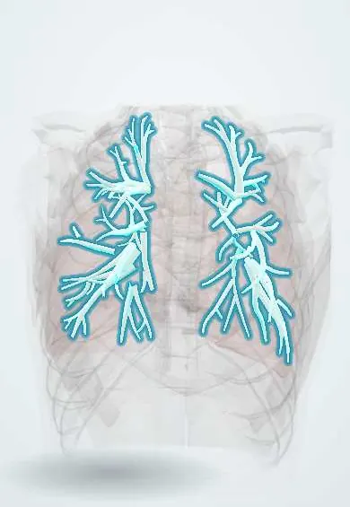
Representative model, for educational purposes only.