
Liver metastases and colorectal liver metastases
The liver is where many cancers spread first. For bowel cancer, liver spread can still be cured by surgery if all of it can be removed safely.
Quick facts
- Key signs
- Often none · Right upper quadrant discomfort
- First line
- Parenchyma-sparing resections
Red flags ↓
No doses: the clinician sets them by the current guideline.
What it is
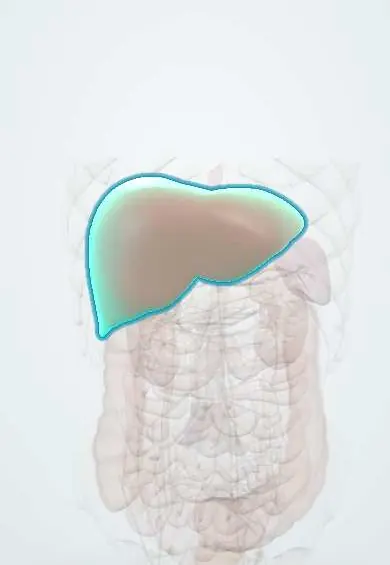
Cancer that has spread to the liver from a primary tumour elsewhere. In most of the world, a malignant lesion found in a non-cirrhotic liver is far more often a metastasis than a primary liver cancer. The commonest sources are the gut organs that drain through the portal vein (colon and rectum, stomach, pancreas, neuroendocrine tumours) and, through the hepatic artery, breast, lung, melanoma (especially uveal) and kidney. Colorectal liver metastases are the classic surgical case: unlike most metastatic disease, they can be cured in selected patients by resection, often combined with chemotherapy.
Who gets it
The liver is the most common site of blood-borne metastases. About half of people with colorectal cancer develop liver metastases, a substantial minority already at diagnosis (synchronous) and the rest later (metachronous). In Colombia colorectal cancer is among the commonest cancers and its incidence is rising, so colorectal liver metastases are a frequent problem for surgical and oncology teams; hepatocellular carcinoma, by contrast, usually arises in cirrhosis.
Causes
- Colorectal cancer (most common and most often treated surgically)
- Other gut primaries draining through the portal vein: stomach, pancreas, gallbladder, small bowel, gastrointestinal neuroendocrine tumours, gastrointestinal stromal tumour
- Breast and lung cancer
- Melanoma, especially uveal melanoma, which characteristically spreads to the liver
- Kidney, ovarian, oesophageal cancer and sarcomas; sometimes an unknown primary
Risk factors
- Advanced primary tumour stage (deep invasion, lymph node spread)
- Raised CEA before or after resection of a colorectal primary
- Biology of the primary: RAS or BRAF mutation in colorectal cancer, high-grade neuroendocrine tumours
- No or incomplete surveillance after treatment of a primary cancer
Pathophysiology
Tumour cells invade vessels in the primary → enter the portal venous (gut) or systemic arterial circulation → are trapped in the liver sinusoids, the first capillary bed for portal blood → survive, attach and grow a new blood supply mainly from the hepatic artery → tumours enlarge, replace liver tissue and may compress bile ducts or vessels → late: jaundice, ascites, liver failure, pain from capsule stretch. Neuroendocrine tumours in the liver release hormones such as serotonin directly into the hepatic veins, bypassing liver breakdown, which is why carcinoid syndrome appears once there are liver metastases.
Classification
- By timing: synchronous (found with the primary) or metachronous (found later)
- By blood supply on imaging: hypovascular (most colorectal, gastric, pancreatic, lung) or hypervascular (neuroendocrine, renal cell, melanoma, thyroid, some breast)
- By resectability: resectable at presentation, potentially resectable after conversion treatment, or unresectable
- By extent: liver-only disease or liver plus extrahepatic disease (lung, peritoneum, nodes)
Symptoms and signs
- Often none: most are found on staging or surveillance scans, or by a rising CEA
- Right upper quadrant discomfort or a dragging pain from stretching of the liver capsule
- Weight loss, fatigue, loss of appetite
- Enlarged, hard, irregular liver on palpation in advanced disease
- Jaundice, ascites and confusion in extensive disease (liver failure or bile duct obstruction)
- Flushing, diarrhoea and wheeze in neuroendocrine tumours (carcinoid syndrome)
Diagnosis
- Clinical
- In a patient with known cancer, a new liver lesion is a metastasis until proven otherwise; in a patient without known cancer, look for the primary (colonoscopy, breast and skin examination, history of eye melanoma) and check for cirrhosis, which points instead to hepatocellular carcinoma. Assess fitness for surgery and the extent of disease outside the liver.
- Tests
- Liver tests (alkaline phosphatase and GGT may rise; normal tests do not exclude metastases), CEA for colorectal cancer (baseline and follow-up), AFP to help separate hepatocellular carcinoma, CA 19-9 and other markers by suspected primary; chromogranin A and urinary 5-HIAA for neuroendocrine tumours. Biopsy is needed when the primary is unknown or the diagnosis is uncertain, but is avoided in a typical, potentially resectable colorectal metastasis because of the small risk of seeding along the needle track. Tumour molecular testing (RAS, BRAF, mismatch repair status) guides systemic therapy.
- Imaging
- Contrast CT of the chest, abdomen and pelvis stages the disease. MRI of the liver with a liver-specific (hepatobiliary) contrast agent and diffusion-weighted imaging is the most sensitive for small lesions and is used to plan surgery. Colorectal metastases are typically hypodense with rim enhancement on the portal venous phase; hypervascular metastases light up in the arterial phase. PET-CT looks for disease outside the liver before major surgery. Contrast-enhanced ultrasound and intraoperative ultrasound help characterise and find small lesions. CT volumetry measures the future liver remnant.
Differential diagnosis
- Hepatocellular carcinoma (cirrhosis, arterial enhancement with washout, raised AFP)
- Haemangioma (peripheral nodular enhancement that fills in)
- Focal nodular hyperplasia and hepatocellular adenoma
- Simple liver cyst or hydatid cyst
- Pyogenic or amoebic liver abscess
- Intrahepatic cholangiocarcinoma
- Focal fatty change or fatty sparing
A liver lesion on the scan: which one?
| Lesion | Typical patient | Imaging clue | Decisive point |
|---|---|---|---|
| Metastasis | Known or hidden primary cancer, often several lesions | Hypodense with rim enhancement (colorectal), or bright in arterial phase (neuroendocrine) | History, primary search, MRI with liver-specific contrast; biopsy if primary unknown |
| Haemangioma | Any adult, often incidental | Peripheral nodular enhancement that fills in towards the centre | Typical MRI pattern; no biopsy needed |
| Focal nodular hyperplasia | Young woman, incidental | Uniform arterial enhancement with a central scar | Retains liver-specific contrast on MRI |
| Hepatocellular carcinoma | Cirrhosis or chronic hepatitis B | Arterial enhancement then washout | Imaging criteria in cirrhosis, AFP |
| Pyogenic abscess | Fever, biliary disease, diabetes | Rim-enhancing collection, sometimes with gas | Aspiration and culture |
| Hydatid cyst | Rural, contact with dogs and sheep | Cyst with daughter cysts or a floating membrane | Serology and typical imaging; never blind puncture |
Goal of treatment
For colorectal (and selected neuroendocrine) metastases: aim for cure by removing or destroying all disease while preserving enough liver. For most other metastases, or when cure is not possible: control the cancer with systemic therapy, relieve symptoms and protect quality of life.
Treatment
- Surgical
- Liver resection for colorectal metastases when all lesions can be removed with clear margins, leaving an adequate future liver remnant (enough healthy liver with blood inflow, outflow and bile drainage) and with no, or controllable, disease outside the liver. Parenchyma-sparing resections are preferred; synchronous primaries can be removed in one or two stages, decided by a multidisciplinary team.
- Medical
- Systemic chemotherapy (fluoropyrimidine-based combinations), around surgery or as conversion therapy to shrink unresectable disease, with targeted agents chosen by tumour biology (anti-EGFR antibodies in RAS wild-type, anti-VEGF agents) and immunotherapy for mismatch repair-deficient tumours. Treatment for metastases from other primaries follows that cancer's own regimens.
- Procedure
- Techniques to make more patients resectable: portal vein embolisation (or combined portal and hepatic vein embolisation) to grow the future remnant, and two-stage hepatectomy. Thermal ablation (radiofrequency or microwave) for small lesions, alone or with resection; stereotactic body radiotherapy when ablation or surgery is not possible.
- Procedure
- Liver-directed treatment for unresectable liver-dominant disease: selective internal radiotherapy (radioembolisation), transarterial chemoembolisation (mainly for neuroendocrine metastases) or hepatic arterial infusion chemotherapy in specialist centres.
- Follow-up
- After treatment: regular CEA and cross-sectional imaging, because recurrence in the liver or lungs is common and may itself be resectable again.
- Conservative
- Palliative care when disease is extensive or the patient is frail: control of pain, itching and ascites, biliary stenting for jaundice, and attention to nutrition and the patient's goals.
Complications
- Liver failure from tumour replacement or after extensive resection (post-hepatectomy liver failure)
- Obstructive jaundice from bile duct compression
- Bile leak, bleeding and infection after resection
- Chemotherapy-associated liver injury (fatty liver, sinusoidal injury) increasing surgical risk
- Disappearing metastases on chemotherapy that still contain cancer cells
- Recurrence in the liver or elsewhere
- Carcinoid crisis during treatment of neuroendocrine metastases
Common traps
- Cancer in the liver means it cannot be cured: for colorectal cancer, complete resection of liver metastases gives long-term survival and cure in a proportion of patients
- More than three or four metastases means unresectable: resectability depends on whether all disease can be removed while leaving enough healthy liver, not on a fixed number of lesions
- Every new liver lesion should be biopsied: in a typical, potentially resectable colorectal metastasis, biopsy adds little and risks seeding; biopsy is for an unknown primary or doubtful diagnosis
- A lesion that disappears on chemotherapy is cured: many still hold cancer cells, so surgeons map lesions before chemotherapy and resect the original sites when possible
- Every malignant liver lesion is hepatocellular carcinoma: in a liver without cirrhosis, a metastasis is far more likely; search for the primary
Prognosis
Depends on the primary. Untreated, liver metastases generally carry a poor outlook, but colorectal liver metastases that are fully resected give long-term survival in a large share of patients, and some are cured; recurrence is common but often treatable. Neuroendocrine liver metastases can have a long course. Metastases from pancreatic, gastric, lung or uveal melanoma primaries are usually treated with systemic therapy and rarely cured.
Prevention
- Colorectal cancer screening, which finds cancers before they spread
- Structured follow-up after colorectal cancer resection (CEA and imaging) to find metastases while still resectable
- Discuss every patient with liver metastases in a multidisciplinary team including a liver surgeon, before deciding they are unresectable
- Genetic assessment for Lynch syndrome and other hereditary cancers
Test yourself
Think of the answer, then tap the question to see it.
A 62-year-old had a sigmoid cancer resected two years ago. His CEA is rising and MRI shows three lesions in the right liver. What is the next step?
Stage for extrahepatic disease (CT chest, often PET-CT) and discuss in a multidisciplinary team with a liver surgeon; if the liver remnant is adequate, resection, often with chemotherapy, can be curative.
Why is the liver the commonest site of metastasis for colorectal cancer?
Venous blood from the colon drains through the portal vein, so the liver sinusoids are the first capillary bed to trap tumour cells shed from the primary.
What decides whether colorectal liver metastases are resectable?
Whether all tumours can be removed with clear margins while leaving an adequate future liver remnant with inflow, outflow and bile drainage, in a fit patient with no uncontrollable disease elsewhere.
Why does carcinoid syndrome appear only once a neuroendocrine tumour of the gut has spread to the liver?
Hormones from a gut primary pass through the portal vein to the liver, which inactivates them; liver metastases release them straight into the hepatic veins and systemic circulation.
The future liver remnant is too small for a planned hepatectomy. What can be done?
Portal vein embolisation of the side to be removed makes the remaining liver grow; a two-stage hepatectomy or combining resection with ablation are other options.
Why it happens
- Metastases to the liver: the most common liver malignancy
- Colorectal adenocarcinoma
- Invasion and metastasis: the metastatic cascade, step by step
- Pathways of spread and the usual metastatic sites
Connections
Educational information, not medical advice. For any symptom, see a health professional.
Sources: Liver Metastasis (StatPearls (NCBI Bookshelf), 2023); Colon Cancer (StatPearls (NCBI Bookshelf), 2025); Liver metastases: MedlinePlus Medical Encyclopedia (MedlinePlus (U.S. National Library of Medicine), 2025).
Spotted a mistake?We are always improving for the community. Tell us what is wrong and our team will review it.Report a mistake
Representative model, for educational purposes only.