Haemothorax !
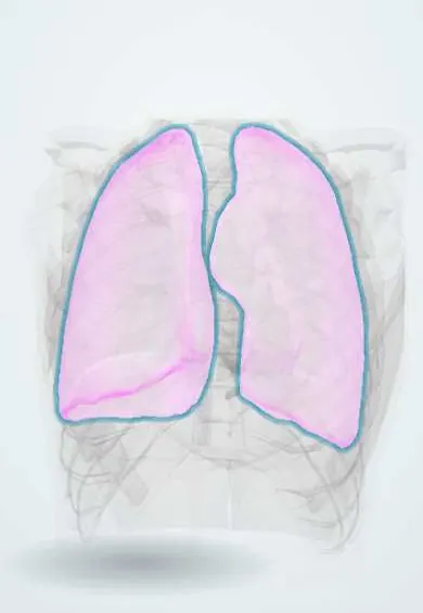
A hemothorax is blood collected between the lung and the chest wall, usually after chest trauma; a large bleed compresses the lung and causes shock.
Updated 2026-10-29
Structures involved
Tap one to see what it is and open it in 3D.
- Pleura
- Posterior intercostal arteries
- Internal thoracic artery
- Intercostal veins
- Ribs (back part)
- Thoracic aorta
In the clinical exam
In physiology
Why it happens
- Hemorrhage: petechiae, purpura, ecchymosis, hematoma, and what blood loss does
- Shock: types, stages and the organs it damages
- Serous membranes: pleura and peritoneum
What it is
Blood in the pleural space, most often after chest trauma. A large amount compresses the lung and causes shock; massive haemothorax is a life-threatening emergency.
Who gets it
Common after both blunt and penetrating chest trauma; in Colombia and Latin America, stab and gunshot wounds are a frequent cause in emergency departments. Non-traumatic haemothorax is rare.
Causes
- Blunt trauma with rib fractures and torn intercostal vessels
- Penetrating trauma: stab and gunshot wounds
- Injury to lung, internal thoracic artery, heart or great vessels
- Iatrogenic: central lines, chest drains, pleural biopsy, thoracic surgery
- Non-traumatic: tumours, anticoagulation, aortic rupture, ruptured vascular malformation
Risk factors
- Chest trauma
- Rib fractures
- Anticoagulant or antiplatelet use
- Bleeding disorders
- Pleural or thoracic procedures
- Pleural or lung tumours
Pathophysiology
Bleeding from intercostal or internal thoracic arteries, lung tissue or great vessels collects in the pleural space, which can hold a large part of the circulating volume. Blood loss causes hypovolaemic shock and the collection compresses the lung. Blood that is not drained clots and organises, causing retained haemothorax, empyema or a trapped lung.
Classification
- Traumatic, iatrogenic or spontaneous
- Small, moderate or large by volume
- Massive haemothorax (large initial drainage or ongoing bleeding; thresholds vary)
- Retained (clotted) haemothorax
Symptoms and signs
- Chest pain and shortness of breath
- Reduced breath sounds on the affected side
- Dullness to percussion
- Tachycardia and hypotension
- Signs of the causative injury
Diagnosis
- Clinical
- Trauma with reduced breath sounds, dullness and shock. In trauma the primary survey and eFAST come first.
- Tests
- Blood count and crossmatch; pleural fluid haematocrit (a value of about half the blood haematocrit or more is the usual definition); coagulation studies.
- Imaging
- Bedside ultrasound (eFAST) detects fluid quickly; upright chest X-ray shows blunting or opacity, while a supine film may show only diffuse haziness; CT for stable patients and retained blood.
Differential diagnosis
- Pleural effusion
- Empyema
- Pneumothorax
- Lung contusion
- Diaphragmatic rupture
Goal of treatment
Restore circulating volume, drain the blood to re-expand the lung, stop ongoing bleeding and prevent retained haemothorax and empyema.
Treatment
- Acute
- Trauma resuscitation with blood products and treatment of coagulopathy.
- Procedure
- Chest tube (tube thoracostomy) to drain the blood, measure losses and re-expand the lung; very small collections may only be watched.
- Surgical
- Thoracotomy or video-assisted surgery for massive haemothorax or ongoing bleeding, using thresholds set by trauma guidelines.
- Procedure
- Early video-assisted thoracoscopic evacuation of retained clotted blood; intrapleural fibrinolytics in selected cases.
- Follow-up
- Repeat imaging, drain output records and watch for fever (empyema).
Complications
- Hypovolaemic shock
- Retained haemothorax
- Empyema
- Fibrothorax and trapped lung
- Respiratory failure
Prognosis
Depends on the source of bleeding and associated injuries. Most patients do well with a chest tube alone; delayed drainage of retained blood increases the risk of empyema and fibrothorax.
Prevention
- Road safety and violence prevention
- Ultrasound guidance for pleural and central line procedures
Test yourself
Think of the answer, then tap the question to see it.
What is the first-line procedure for a traumatic haemothorax?
A chest tube (tube thoracostomy) to drain the blood, re-expand the lung and monitor ongoing bleeding.
When does a haemothorax usually need surgery?
When it is massive on drainage or bleeding continues, by thresholds set in trauma guidelines, or when clotted blood is retained.
How is haemothorax defined from pleural fluid?
By a pleural fluid haematocrit of about half or more of the blood haematocrit.
What can happen if a haemothorax is not fully drained?
Retained clot can become infected (empyema) or organise into a fibrothorax that traps the lung.
Related conditions
- Rib fractures
- Pneumothorax
- Pleural effusion
- Hypovolaemic shock
- Aortic dissection
- Flail chest
- Cardiac tamponade
Educational information, not medical advice. For any symptom, see a health professional.
Sources: Hemothorax (StatPearls (NCBI Bookshelf), 2023).
Spotted a mistake?We are always improving for the community. Tell us what is wrong and our team will review it.Report a mistake
Representative model, for educational purposes only.