
Air embolism (venous and arterial gas embolism)
Air that gets into a vein or artery, usually through a central line, a procedure or an injury, can block the heart or the brain's blood flow within seconds.
Quick facts
- Key signs
- Sudden breathlessness · Sudden hypotension · Seizures
- First line
- Hyperbaric oxygen when there are neurological deficits
Red flags ↓
No doses: the clinician sets them by the current guideline.
What it is
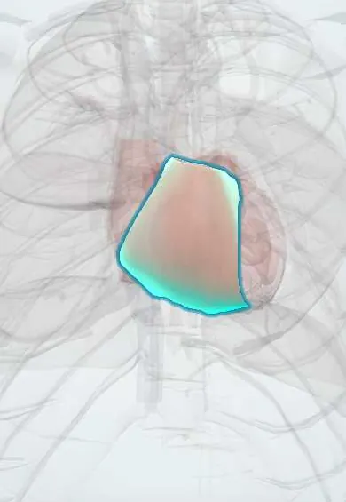
Entry of gas into the bloodstream through an opening between air and a vessel plus a pressure gradient that drives it in. Venous air embolism travels to the right heart and lungs; arterial gas embolism reaches organs such as the brain and heart, either directly or across a patent foramen ovale. The effect depends on the volume, the speed of entry and the route.
Who gets it
Rare and under-reported. In one large series of central venous catheter placements it occurred in about 1 in 772. Small, harmless bubbles from peripheral lines are seen on up to 23% of chest CT scans. The main causes have shifted from open surgery and trauma towards endoscopy, angiography, tissue biopsy and vascular access. Even with treatment, one-year mortality in recent series is around 21%.
Causes
- Central venous catheters: insertion, disconnection and especially removal
- Angiography and other endovascular or interventional procedures
- Surgery with the operative site above the heart (neurosurgery in the sitting position, cardiac surgery)
- CT-guided lung biopsy (air entering a pulmonary vein)
- Penetrating chest trauma and lung barotrauma, including diving
- Peripheral intravenous lines and infusion tubing with air
Risk factors
- Low central venous pressure: hypovolaemia, upright or sitting position
- Deep inspiration (negative intrathoracic pressure) while a central vein is open
- Short skin-to-vein tracts and large sheaths
- Patent foramen ovale or septal defect (venous air can cross to the arteries)
- Unprimed tubing, loose connections and open catheter hubs
Pathophysiology
Open vessel + pressure gradient → air enters. Venous route: central venous pressure is often below atmospheric pressure, especially in inspiration, hypovolaemia or sitting up → air is sucked in → a large bolus lodges in the right ventricular outflow tract as an 'air lock' → pulmonary artery pressure rises → right heart strain, falling cardiac output and circulatory collapse; smaller volumes reach the lungs and are absorbed. Arterial route: air injected into an artery, or venous air crossing a patent foramen ovale (paradoxical embolism) → bubbles block end arteries in the brain or heart → ischaemia or infarction even with small volumes.
Classification
- Venous air embolism: right heart and pulmonary circulation
- Arterial gas embolism: direct (angiography, lung biopsy, lung barotrauma) or paradoxical through a patent foramen ovale
- By setting: iatrogenic (lines, procedures, surgery), traumatic, and diving-related (see decompression sickness)
Symptoms and signs
- Sudden breathlessness, continuous coughing and chest pain
- Sudden hypotension, tachycardia, arrhythmia or cardiac arrest
- Seizures, loss of consciousness, confusion or one-sided weakness (arterial or paradoxical)
- During anaesthesia: a sudden fall in end-tidal carbon dioxide, often the earliest sign
- Falling oxygen saturation, usually a late sign
Diagnosis
- Clinical
- Mostly clinical: sudden cardiorespiratory or neurological deterioration at the moment a vessel is open to air. It is often missed and labelled a thrombotic stroke, so the timing next to a procedure is the clue. Treatment starts on suspicion.
- Tests
- ECG (tachycardia, ST changes, right heart strain); arterial blood gases (hypoxaemia and hypercarbia); transoesophageal echocardiography detects very small volumes of intracardiac air; end-tidal carbon dioxide monitoring under anaesthesia.
- Imaging
- CT of the brain, chest or heart can show intravascular air, which may clear within hours; brain MRI shows the resulting infarcts.
Differential diagnosis
- Pulmonary thromboembolism
- Ischaemic stroke from a thrombus
- Tension pneumothorax after line insertion
- Anaphylaxis or vasovagal collapse during a procedure
- Fat or amniotic fluid embolism
- Decompression sickness in divers
Venous, arterial and diving gas embolism compared
| Type | How the air gets in | What it blocks | Typical picture | Position and key treatment |
|---|---|---|---|---|
| Venous air embolism | Open central vein with low venous pressure: line insertion or removal, surgery above the heart | Right ventricular outflow tract and pulmonary circulation | Sudden breathlessness, hypotension, falling end-tidal carbon dioxide, collapse | Left side down and head down; oxygen; aspirate through a right heart catheter if present |
| Arterial gas embolism (iatrogenic or paradoxical) | Air injected in an artery, lung biopsy, or venous air crossing a patent foramen ovale | Brain and coronary end arteries | Seizure, coma, aphasia or weakness during or just after the procedure | Flat on the back, not head-down; oxygen; hyperbaric oxygen for neurological deficits |
| Diving arterial gas embolism | Lung overexpansion on ascent pushes alveolar gas into pulmonary veins | Brain arteries | Stroke-like signs within minutes of surfacing | Flat, oxygen and recompression in a hyperbaric chamber |
Goal of treatment
Stop more air entering, keep the circulation going, shrink the bubbles with oxygen and pressure, and limit brain and heart injury.
Treatment
- Acute
- Stop the entry: close or clamp the line, cover the site with an occlusive dressing; in surgery, compress the jugular veins and seal the entry site; ABC, cardiopulmonary resuscitation if needed and call for help.
- Acute
- Venous air embolism: Durant manoeuvre, left side down and head down, to move air out of the right ventricular outflow tract into the right atrium. Arterial gas embolism: keep the patient flat on the back, because head-down may worsen cerebral oedema.
- Medical
- The highest available concentration of oxygen through a non-rebreather mask or the ventilator: it oxygenates ischaemic tissue and shrinks bubbles by washing nitrogen out of them.
- Procedure
- Aspiration of air through a catheter already in the right atrium or ventricle when there is an air lock with collapse; extracorporeal support in selected cases.
- Procedure
- Hyperbaric oxygen when there are neurological deficits: it is the treatment of choice and works best when started early, ideally within hours.
- Follow-up
- Intensive care with haemodynamic and neurological monitoring after stabilisation.
Complications
- Right heart failure and cardiac arrest
- Stroke, seizures and permanent neurological deficits
- Myocardial ischaemia from coronary air
- Death
Common traps
- Every gas embolism goes head-down and left side down: that position is for venous air; in arterial gas embolism the patient lies flat because head-down may worsen cerebral oedema
- Oxygen is needed only if the saturation falls: oxygen also shrinks the bubbles, and desaturation is a late sign, so the highest concentration is given at once
- Removing a central line is safer than inserting it: removal opens a tract to a low-pressure vein, so the patient lies flat or head-down and the site is sealed with an occlusive dressing
- Venous air cannot reach the brain: through a patent foramen ovale it can cross to the arteries and cause a stroke
- Sudden deficits after a procedure are a thrombotic stroke: air embolism is often mislabelled this way; the timing next to a line, biopsy or angiogram is the clue
Prognosis
Small venous bubbles are usually absorbed without harm, but large venous boluses and arterial emboli can kill or disable within minutes. Hyperbaric oxygen started early gives the best outcomes, and late treatment may still help.
Prevention
- Insert central lines with the patient head-down and well hydrated, avoiding entry during inspiration; occlude open hubs and flush all lumens
- Remove central lines with the patient flat or head-down and the site below the heart, during a Valsalva manoeuvre or active expiration
- Cover the removal site with an occlusive dressing, apply pressure and keep the patient lying down afterwards
- Locking connectors, primed tubing, air-free syringes and air-in-line detectors on pumps
- Monitoring end-tidal carbon dioxide and, in high-risk surgery, Doppler or transoesophageal echocardiography
Test yourself
Think of the answer, then tap the question to see it.
A patient sitting up in bed becomes breathless and hypotensive the moment his jugular line is pulled out. What happened, and why did sitting up matter?
Venous air embolism: with the patient upright, central venous pressure is below atmospheric, so air was sucked through the open tract into the right heart.
How is the patient positioned for a venous air embolism, and why?
Left side down and head down (Durant manoeuvre), so the bubble floats out of the right ventricular outflow tract into the right atrium and the air lock is relieved.
After a cerebral angiogram a woman has sudden aphasia and arm weakness. Why should she not be tipped head-down?
This is arterial gas embolism to the brain; head-down may worsen cerebral oedema, so she lies flat on the back with high-concentration oxygen and hyperbaric oxygen is considered.
How can air from a peripheral vein cause a stroke?
Through a patent foramen ovale or septal defect, venous air can cross from the right to the left heart and embolise to the brain (paradoxical embolism).
Why is oxygen given at the highest concentration even if the saturation is normal?
Oxygen washes nitrogen out of the bubbles and shrinks them, and supports ischaemic tissue; a falling saturation is a late sign.
Connections
Educational information, not medical advice. For any symptom, see a health professional.
Sources: Air Embolism: Practical Tips for Prevention and Treatment (Journal of Clinical Medicine (PubMed Central), 2016); Prevention of air embolism related to central venous catheter removal: an integrative review (Jornal Vascular Brasileiro (PubMed Central), 2025); Anesthesiology Considerations and Management of Venous Air Embolism in Patients in the Semisitting Position: A Single-Center Review (Cureus (PubMed Central), 2025).
Spotted a mistake?We are always improving for the community. Tell us what is wrong and our team will review it.Report a mistake
Representative model, for educational purposes only.