
Upper tract urothelial carcinoma (renal pelvis and ureter)
A cancer of the lining of the kidney's collecting system or the ureter. It usually shows up as blood in the urine and often returns later in the bladder.
Quick facts
- Key signs
- Visible haematuria · Flank or back pain · Tiredness
- Diagnosis
- Ureteroscopy with biopsy and selective cytology
Red flags ↓
No doses: the clinician sets them by the current guideline.
What it is
A malignant tumour of the urothelium lining the renal calyces, renal pelvis and ureter. It is the same cell type as bladder cancer but much rarer (about 5–10% of urothelial cancers), more often invasive at diagnosis, and treated by removing the kidney, ureter and a cuff of bladder unless the tumour is low risk.
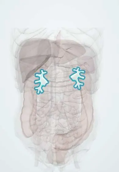
See it in 3D
Who gets it
Rare: urothelial cancers of the upper tract make up roughly 5–10% of all urothelial carcinomas, with an incidence of about one to two per 100 000 a year in Western countries. Men are affected about three times as often as women, with a peak at 70–90 years. Tumours of the renal pelvis are about twice as common as those of the ureter. About 60% are already muscle-invasive at diagnosis, compared with 15–25% of bladder cancers. In Taiwan they make up 20–25% of urothelial cancers, linked to aristolochic acid.
Causes
- Tobacco smoking, the most common risk factor, shared with bladder cancer
- Occupational aromatic amines (dye, rubber, leather and textile industries)
- Aristolochic acid from Aristolochia plants: Balkan endemic nephropathy (contaminated flour) and herbal remedies; it leaves a signature TP53 mutation
- Lynch syndrome (germline DNA mismatch repair defect, most often MSH2): upper tract urothelial cancer is its third most common cancer after colon and endometrium
- Heavy long-term use of phenacetin-containing analgesics; arsenic in drinking water
- Chronic stones and infection (linked to the rarer squamous cell type)
Risk factors
- Current or past smoking
- Previous or current bladder cancer
- Personal or family history of colorectal or endometrial cancer (possible Lynch syndrome)
- Use of traditional herbal medicines that may contain Aristolochia, or residence in endemic Balkan areas
- Occupational exposure to aromatic amines
- Older age and male sex
Pathophysiology
A carcinogen excreted in urine (tobacco metabolites, aromatic amines, aristolochic acid adducts) or an inherited defect in DNA mismatch repair → mutations accumulate in urothelial cells across the whole lining (field change) → tumours arise in the renal pelvis or ureter, often several at once → low-grade tumours tend to grow as papillary fronds into the lumen, high-grade tumours invade early because the wall of the pelvis and ureter is thin and the muscle layer is close → a tumour that blocks the lumen causes hydronephrosis and flank pain, and one that bleeds causes haematuria → tumour cells shed downstream seed the bladder, so 30–50% of patients later develop bladder tumours. Aristolochic acid concentrates its DNA damage in the kidney and upper tract, which explains multiple and bilateral tumours with relatively few in the bladder.
Classification
- By site: renal pelvis and calyces, or ureter (distal ureter most often)
- By grade: low grade or high grade (the strongest guide to behaviour together with stage)
- By stage (TNM): Ta and carcinoma in situ (non-invasive), T1 (connective tissue), T2 (muscle), T3 (beyond muscle into renal parenchyma or fat), T4 (adjacent organs); then nodes and metastases
- By risk for treatment: low risk (single, small, low-grade, non-invasive on imaging) or high risk (hydronephrosis, large, high-grade, multifocal, variant histology or previous cystectomy)
- Histology: urothelial (transitional cell) carcinoma in the great majority; squamous cell carcinoma is uncommon and tied to stones and infection
Symptoms and signs
- Visible haematuria, often painless; the most common presentation
- Flank or back pain, sometimes colicky when clots or the tumour block the ureter
- Tiredness, weight loss or bone pain in advanced disease
- Found incidentally on imaging done for other reasons
Diagnosis
- Clinical
- Ask about smoking, occupation, herbal remedies and a family history of colon, endometrial or upper tract cancer. Screen every patient for Lynch syndrome features; genetic referral is advised for young patients and those with a personal or family history of Lynch-spectrum cancers.
- Tests
- Urinalysis and urine cytology (sensitive for high-grade tumours, poor for low-grade). Cystoscopy to look for coexisting bladder tumours. Ureteroscopy with biopsy and selective cytology confirms the diagnosis and grade. Tumour testing for mismatch repair deficiency (immunohistochemistry or microsatellite instability) when Lynch syndrome is suspected. Kidney function, because surgery removes one kidney.
- Imaging
- CT urography is the main imaging test: a filling defect, wall thickening or hydronephrosis, plus staging of nodes and organs. MR urography when contrast CT is not possible. Chest imaging, and bone imaging if symptoms suggest spread.
Differential diagnosis
- Bladder cancer (same symptoms; cystoscopy and upper tract imaging separate them, and both can coexist)
- Renal cell carcinoma (solid parenchymal mass rather than a collecting-system defect)
- Radiolucent urinary stone or blood clot (filling defect without tissue on ureteroscopy)
- Renal papillary necrosis (sloughed papilla in the collecting system)
- Benign ureteral polyp or stricture
Haematuria with a lesion in the kidney or ureter: which one is it?
| Lesion | Where it sits | Typical clues | What settles it |
|---|---|---|---|
| Upper tract urothelial carcinoma | Lining of calyces, renal pelvis or ureter | Smoker, older man; positive cytology if high grade; may have had bladder cancer | CT urography filling defect, ureteroscopy and biopsy |
| Renal cell carcinoma | Kidney parenchyma (cortex) | Solid enhancing mass that bulges the kidney outline; cytology negative | Contrast CT or MRI of the kidney; biopsy rarely needed |
| Bladder cancer | Bladder wall | Same risk factors and haematuria; irritative voiding symptoms | Cystoscopy and biopsy; always image the upper tract too |
| Urinary stone or clot | Inside the collecting system or ureter | Colicky pain; stone dense on non-contrast CT, clot changes over time | Non-contrast CT; ureteroscopy if a defect persists |
Goal of treatment
Remove the cancer while keeping as much kidney function as is safe, choosing kidney-sparing treatment for low-risk tumours and radical surgery for high-risk ones, and catch the frequent bladder recurrences early.
Treatment
- Surgical
- Radical nephroureterectomy with excision of a bladder cuff around the ureteric orifice (open, laparoscopic or robot-assisted) is the standard for high-risk tumours; leaving the distal ureter behind risks recurrence in that stump.
- Procedure
- Kidney-sparing treatment for low-risk tumours: ureteroscopic or percutaneous laser ablation or resection, or segmental resection of the distal ureter with reimplantation into the bladder. It needs a patient willing to have repeated ureteroscopy and imaging. Also considered for selected high-risk tumours in a solitary kidney or severe chronic kidney disease.
- Medical
- Platinum-based chemotherapy around surgery for invasive disease; it is often given before nephroureterectomy because kidney function is better while both kidneys remain. Chemotherapy and immune checkpoint inhibitors for metastatic disease. Topical instillation into the upper tract after kidney-sparing surgery is used in some centres, with limited evidence.
- Follow-up
- Lifelong surveillance with cystoscopy and urine cytology for bladder recurrence, plus CT urography of the remaining upper tract; after kidney-sparing treatment, repeat ureteroscopy of the treated side. Patients with Lynch syndrome and their relatives are referred for genetic counselling and colon and endometrial screening.
Complications
- Recurrence in the bladder (30–50% of patients)
- Recurrence in the opposite upper tract (a few per cent; more after aristolochic acid exposure)
- Chronic kidney disease after removing one kidney
- Hydronephrosis and loss of function of the affected kidney
- Spread to lymph nodes, lung, liver and bone
Common traps
- Haematuria explained by a normal cystoscopy: the bladder is only part of the urothelium, so visible haematuria needs imaging of the kidneys and ureters as well
- Removing the kidney and most of the ureter is enough: the intramural ureter and its orifice are a common site of recurrence, so a cuff of bladder is taken with them
- Urine cytology rules it out: cytology catches high-grade tumours but often misses low-grade ones, which need ureteroscopy
- Upper tract cancer is just bladder cancer in another place: it is invasive at diagnosis far more often, and it can be the first sign of Lynch syndrome in the patient's family
- Herbal teas are harmless: some traditional remedies contain aristolochic acid, which destroys kidney tissue and causes upper tract cancers years after the exposure stops
Prognosis
Depends mainly on stage and grade. Non-invasive or superficial tumours are cured in more than 90% of cases, while deeply invasive tumours have a much poorer outlook, and metastatic disease is rarely curable. About three quarters of patients are alive five years after radical nephroureterectomy in large series. Tumours arising in Lynch syndrome tend to do better than sporadic ones at the same stage.
Prevention
- Not smoking, and stopping at any age
- Avoiding herbal products that may contain Aristolochia; workplace protection from aromatic amines
- Genetic counselling and urinalysis-based follow-up for people with Lynch syndrome
- Prompt imaging of the whole urinary tract for visible haematuria
Test yourself
Think of the answer, then tap the question to see it.
Why is a cuff of bladder removed together with the kidney and ureter in radical nephroureterectomy?
The intramural ureter and its orifice are a frequent site of recurrence and are hard to watch by imaging or endoscopy, so they are taken with the specimen.
Which inherited syndrome should you consider in a 50-year-old with upper tract urothelial cancer and a mother with endometrial cancer?
Lynch syndrome (germline mismatch repair defect, most often MSH2); upper tract urothelial cancer is its third most common cancer.
What is the most frequent site of new tumours after treatment of an upper tract urothelial carcinoma?
The bladder: 30–50% of patients develop bladder tumours, so lifelong cystoscopy is needed.
Which low-risk features allow kidney-sparing treatment?
A single, small, low-grade tumour on cytology and biopsy with no sign of invasion on CT urography, in a patient willing to have close endoscopic follow-up.
Why is chemotherapy often preferred before rather than after nephroureterectomy?
Platinum drugs need good kidney function, which is better while both kidneys are still present; after removing one, many patients no longer qualify.
Why it happens
Connections
Educational information, not medical advice. For any symptom, see a health professional.
Sources: Transitional Cell Cancer of the Renal Pelvis and Ureter Treatment (PDQ), patient version (National Cancer Institute, NIH, 2026); Transitional Cell Cancer of the Renal Pelvis and Ureter Treatment (PDQ), health professional version (National Cancer Institute, NIH, 2026); Optimal management of upper tract urothelial carcinoma: current perspectives (Leow et al., OncoTargets and Therapy) (Europe PMC, 2020); Lynch syndrome and exposure to aristolochic acid in upper-tract urothelial carcinoma: its clinical impact? (Colin et al., Translational Andrology and Urology) (Europe PMC, 2016).
Spotted a mistake?We are always improving for the community. Tell us what is wrong and our team will review it.Report a mistake
Representative model, for educational purposes only.